Research: Singapore researchers find that dressing fibres aligned in parallel help wounds heal faster
 | By Assistant Professor Navin Verma, Assistant Professor of Immunology and Cell Biology at LKCMedicine
|
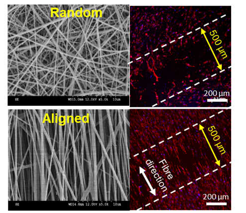
An interdisciplinary team of researchers from Nanyang Technological University Singapore’s (NTU) LKCMedicine, the Singapore Eye Research Institute (SERI), and the National University of Singapore (NUS) has developed a skin-mimicking scaffold by a technique, called “electrospinning”. Using this technique and a composition of polycaprolactone (PCL) and gelatin, they fabricated nanofibers in parallel alignment. They found that this skin-mimicking scaffold with fibres aligned in parallel helps wounds heal significantly faster than fibres with random alignment.
When skin is injured or wounded, it takes some time to heal. This depends on how deep or large the injury cut is. Certain types of skin wounds, such chronic wounds, usually fail to heal, may take months or even years to heal, or may recur after initial healing. The wound healing process in an individual is delayed if the patient has underlying medical conditions such as diabetes, a compromised immune system, skin cancers, or infections. Delayed wound healing further adds to the suffering of patients and their well-being, requiring special treatment and care and adding to the health burdens in wound care.
With the goal of speeding up the process of wound healing, researchers used a simple electrospinning method to prepare a novel composite dressing. They mixed two biodegradable materials - a polyester PCL and gelatin, a protein product. To increase the mechanical strength and durability of the dressing materials, they cross-linked the fibres using a bio-adhesive chemical, dopamine hydrochloride. Scaffolds made from these fibres were hydrophilic and mimicked the structure and properties of the natural skin, which is important for the skin cells to adhere and move.
Enhanced wound healing of skin cells in vitro
“We tuned fibre alignment by adjusting the speed of the rotating drum collector of the electrospinning system,” said Mr Erfan Rezvani Ghomi, a PhD student at NUS and the first author of the paper. “The hydrophilic fibres would absorb excessive fluid and create a moist environment when applied on the wound, which is a desirable feature of a dressing.”
To prevent bacterial colonisation on the wound, researchers incorporated a naturally occurring anti-infective ingredient, called ε-polylysine, to the nanofibrous scaffolds. In separate experiments conducted previously, researchers have demonstrated that ε-polylysine possess broad-spectrum antimicrobial properties and is skin-compatible.
“We leveraged our experience in designing antimicrobial nanofibers. The PCL/Gelatin scaffolds containing ε-polylysine prevented bacterial growth and enhanced wound healing,” said Dr Rajamani Lakshminarayanan, one of the lead Principal Investigators (PIs) of the study at SERI.
The research team experimented with various topologies of the nanofibers using the cell culture system. Scaffolds with fibres arranged in parallel showed increased cell proliferation and accelerated cell movement in the direction of the fibre alignment in comparison to that with fibres arranged in random.

LKCMedicine Assistant Professor Navin Verma, one of the lead PIs of the study
“Our findings that PCL/gelatin scaffolds with parallel fibres increase cell proliferation and migration is interesting. This study lays the groundwork for new approaches in dealing with wound healing. Our next step is to add anti-inflammatory functionalities to this highly aligned fibre material and investigate how tuning scaffold fibre alignment could also be employed to control inflammation in the wounds,” said Assistant Professor Navin Verma, one of the lead PIs of the study at LKCMedicine.
“Wound care is a multibillion-dollar healthcare problem worldwide and therefore wound care industries are actively exploring this kind of innovative idea. We are beginning to explore such opportunities,” said Professor Seeram Ramakrishna, the head of the Centre for Nanofibers and Nanotechnology at NUS.
Although a range of dressings based on nanofibers and hydrogels of varying composition have been developed and tested for wound healing. But whether structure properties and topologies of the fibrous scaffolds could influence cell behaviours is less studied.
Highlighting the significance of the work, Professor S. C. Kundu, Research Coordinator, Research Institute on Biomaterials, Biodegradables and Biomimetics (3Bs) at the University of Minho, Portugal, who was not involved in the study, said, “Their study offers a general framework for future research that would benefit wound patients. Rapid wound healing is particularly desired in the case of chronic wounds or severe burns. But more research needs to be done.”
The study was published in the highly reputable journal Advance Fibre Materials in October. The paper is available here.