Research: Breakthrough in determining structure of tiny particles at nanometre scale leading to new drug discovery
 | By Sarah Zulkifli, Science Writer, Communications & Outreach |
Every move we make and every pain we feel is caused by a series of electrical signals made by our nerves. These signals are made possible by ion channels – so tiny that the structure had been extremely difficult to determine.
Published in Proceedings of the National Academy of Sciences (PNAS), a team led by Assistant Professor of NTU School of Biological Sciences, Shashi Bhushan’s group in collaboration with Dr Ong Seow Theng, Assistant Director and Centre Lead for LKCMedicine-ICESing Ion Channel Platform (LICP), with Visiting Professor at LKCMedicine, George Chandy, determined the structure of a particular ion channel at near-atomic state – a first in Singapore.
 NTU School of Biological Sciences Assistant Professor Shashi Bhushan
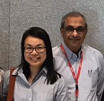 Assistant Director and Centre Lead for LKCMedicine-ICESing Ion Channel Platform Dr Ong Seow Theng (left), and LKCMedicine Visiting Professor George Chandy
A nerve is a bundle of fibres composed of neurons that uses electrical and chemical signals to transmit sensory and motor information from one part of the body to another. The rhythmic beating of the heart and the flexing of muscles and the digestion of our food are all made possible with the help of networks of ion channels. Ion channels are specialised proteins in the membrane that provide a passageway for charged ions to pass through.
When these ion channels become dysfunctional or mutated, it causes episodes of epilepsy, cardiac arrythmias and more. To treat this, drugs that target ion channels are prescribed and are normally used for patients suffering from hypertension, heart diseases, diabetes and more.
In order to understand how these ion channels work, it is vital to determine their structure. Using state-of-the-art cryogenic electron microscopy facilities situated at NTU and the National University of Singapore (NUS), the team determined the structure of a voltage-gated potassium ion channel Kv1.3 that is highly expressed in human immune cells.
Potassium ion channels are proteins with a central canal through which potassium ions flow in and out of cells. The canal has a gate that opens to allow the flow of potassium ions. Flow is rapidly curtailed by a process called slow inactivation because unrestrained flow is harmful. Mutations that affect this process are deleterious.
Asst Prof Bhushan said, “Out of the 78 potassium channels in humans, my group together with Prof Chandy and Dr Ong are studying the structures of two potassium channels at present, the voltage-gated Kv1.3 channel and the calcium-activated KCa3.1. channel. Both are vitally important for coupling external stimuli with intracellular signalling cascades essential for homeostasis and activation in diverse non-excitable cells.”
The Kv1.3 ion channel has been identified to be a target for the cause of diseases that are suffered by much of the population such as psoriasis, multiple sclerosis, Alzheimer’s disease, Parkinson’s disease and leukemia.
The impact of this study is endless: one of which is the design of new drugs. Prof Chandy added, “At least two Kv1.3 blockers are in human trials for the treatment of autoimmune and neuroinflammatory diseases. One of these, dalazatide, was developed by me. One KCa3.1 blocking drug is in human trials for the treatment of Alzheimer's disease and for severe congenital hemolytic anemias.
“We have developed a second KCa3.1 inhibitor, NTU-103, which NTU recently patented. Drugs that target membrane proteins account for a vast number in the pharmacopeia and many are included in the World Health Organization's list of essential medicines.”
The team used advanced technology called cryogenic-electron microscopy (cryo-EM) at facility laboratories located in NTU and NUS. It is a game-changing technology that enables proteins to be probed in unprecedented detail. Flash-frozen samples of proteins are bombarded with electrons. Analysis of images of electrons ricocheting after collision with the sample enables proteins to be seen in sharp detail or at high-resolution.

Cryo-EM density map of Kv1.3-Kvβ2 with a fitted model viewed from the membrane plane (centre), from inside the cell (left), and from the extracellular side (right)
Asst Prof Bhushan said, “Using state-of-the-art facilities at my cryo-EM lab in NTU, we were able to achieve this breakthrough. We defined the functional role of the channel in immune cells, demonstrated its importance as a therapeutic target for autoimmune and neuroinflammatory diseases, and showed its specificity towards inhibitors for the treatment of multiple sclerosis and psoriasis.”
Dr Ong said, “With the recent technology advances in cryo-EM, ion channels can now be studied at nanometre scale. The availability of the atomic-resolution structures would revolutionise the study of ion channels. Ion channel gating mechanism and function can now be better elucidated by structural insights. Understanding of channelopathies – diseased ion channels – caused by mutation-related conformational changes would also be possible.”
Prof Chandy added, “We have now seen this channel as an electrical current, a DNA sequence, a protein sequence, and a high-resolution structure bound to a drug I developed. It has been an enjoyable journey.”
Dr Anu Tyagi, the paper’s first author said, “The lessons learned from this study will have great implications on the perceived mechanism of voltage-gated potassium channels.”
Additionally, a peptide called dalazatide derived from venom released by tentacles of a sea anemone can substantially change the ion channel’s structure. This may inform scientists to study venom-based therapeutics targeting potassium ion channels.
Dr Ong added, “Structural insights at the near-atomic resolution of Kv1.3 could fast-track drug discovery for many human diseases. One example is to guide drug design by turning venomous toxins into useful therapeutics.”
“These venoms are both friend and foe. They have been used for millennia in traditional medicine in many cultures to treat diverse ailments. But excessive activity by these substances can be deleterious,” Asst Prof Bhushan informed.
Moving forward, the team is interested in determining if other peptides, for example from scorpions, will cause the same dramatic change in the structure of the ion channel.
LKCMedicine Professor of Neuroscience and Mental Health George Augustine, who is not part of the study, commented on its impact, “This paper represents the pinnacle of the team’s work at NTU. Aside from showing how a therapeutically-important drug blocks ion conduction through a potassium channel, it also provides some novel insights into how the potassium channel works in the absence of a drug.
“It is the first atomic-level K channel structure solved in Singapore and is the culmination of decades of work that George has done to identify this ion channel and its importance for the immune system.”
Asst Prof Bhushan added, “It would also encourage structural studies in Singapore on other biologically and medically relevant membrane proteins.
“Atomic-resolution of drug-bound structures allow us to peek into drug binding sites and their mechanism of action. Integration of atomic-scale structures and artificial-intelligent-based drug design would accelerate drug discovery in future drug development.”