NTU scientists outsmart resistant bacteria
Research at NTU is paving the way for new treatments that target resistant bacteria.
Antimicrobial resistance is one of the world’s top public health threats. Often termed a silent pandemic, bacteria that are resistant to antimicrobials have caused millions of deaths annually. If left unchecked, the number of deaths attributed to antimicrobial-resistant bacteria is expected to increase to 10 million a year by 2050, according to estimates by the World Health Organisation.
NTU scientists are pioneering strategies to stay one step ahead of these bacteria.
One NTU research team developed a compound that inhibits an enzyme crucial for energy generation in a resistant bacterium.
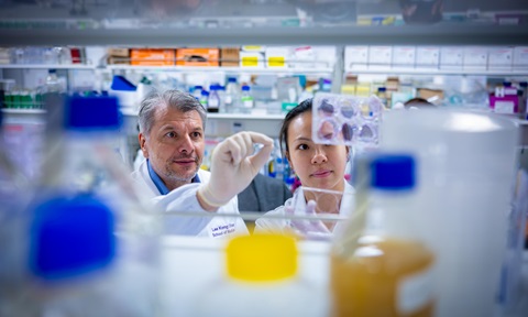
Collaborating with the Agency for Science, Technology and Research in Singapore (A*STAR), another group of researchers from NTU’s Lee Kong Chian School of Medicine (LKCMedicine) uncovered how the same bacterium resists infection by viruses.
To understand how another resistant bacterium colonises its host, a research team from NTU and Imperial College London investigated the molecular structure of the ‘weapon’ it uses to load and fire toxins into rival bacteria and other cells.
Their research has opened avenues for new treatments that target resistant bacteria as well as novel ways of disarming such bacteria.
Blocking energy generation
Opportunistic pathogens that are resistant to antimicrobials are especially concerning as they infect patients with weakened immune systems. For instance, Mycobacterium abscessus causes severe lung disease in patients with cystic fibrosis and is intrinsically resistant to many commonly-used antibiotics.
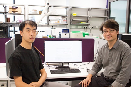
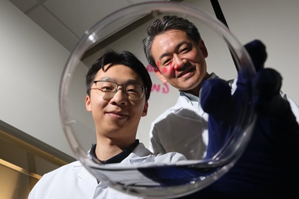
To treat M. abscessus infections, a team of researchers led by Prof Gerhard Grüber of NTU’s School of Biological Sciences (SBS) has developed a compound that prevents the bacterium from generating energy for survival. The compound inhibits a key enzyme in the electron transport chain of M. abscessus, a series of proteins that the bacterium uses to produce adenosine triphosphate (ATP), a molecule that cells use to power their metabolic processes.
The findings were published in Nature Communications.
Using cryo-electron microscopy, the scientists identified a pocket in the cytochrome b subunit of the cytochrome bcc oxidase, a crucial enzyme in the electron transport chain of M. abscessus, that binds to the substrate and is responsible for the activity of the enzyme.
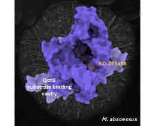
The compound (ND-011458) binds to a cavity in the cytochrome b subunit of the cytochrome bcc oxidase enzyme and inhibits it. Credit: NTU Singapore.
They then designed a compound that fits into the pocket to inhibit the enzyme.
As the structure of the cytochrome bcc oxidase is unique to M. abscessus, the compound only targets the bacterium and is not toxic to human cells.
When used in combination with clofazimine, an antibiotic used to treat mycobacterial infections, the new compound was effective at killing M. abscessus (2-log fold reduction in four days).
“As the currency of life, ATP delivers the energy for essential processes in M. abscessus, including its defence mechanisms against antibiotics. Silencing the electron transport chain that produces ATP is thus a potential treatment for difficult-to-treat M. abscessus infections that also disables the bacterium,” says Prof Gerhard Grüber, who was the corresponding author of the research.
A patent has been filed and the researchers are working with US-based pharmaceutical company Hsiri Therapeutics to license the compound.
Hijacking mycobacteria with viruses
In addition to developing new antimicrobials, viruses called bacteriophages have also been explored as a possible treatment for M. abscessus infections.
These bacteriophages infect the bacteria and hijack their replication machinery to make more copies of themselves. Eventually, the bacteria burst, releasing the newly-formed viruses.
In another study reported in the Proceedings of the National Academy of Sciences, researchers from NTU and the Agency for Science, Technology and Research in Singapore have uncovered how M. abscessus can resist attack by these viruses.
Their findings shed light on how phage therapies can be optimised to treat M. abscessus infections effectively while minimising the development of resistance.
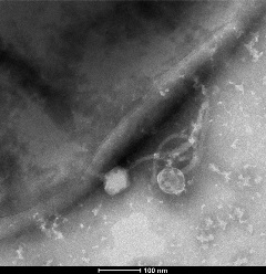
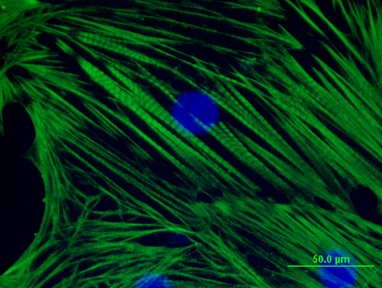
Transmission electron micrograph of a bacteriophage attached to a M. abscessus cell. Credit: NTU Singapore.
M. abscessus exists as two variants: smooth and rough. The smooth variant of M. abscessus produces lipids called glycopeptidolipids on its outer surface and forms round colonies while the rough variant lacks glycopeptidolipids and forms irregular cauliflower-shaped colonies.
Compared to the smooth variant, the rough variant of M. abscessus causes more severe disease and is more challenging to treat.
The researchers discovered the mechanism by treating “smooth” M. abscessus strains with bacteriophages that target them. After treatment, the researchers observed that rough variants of the bacteria emerged and found mutations in the genes coding for essential enzymes required for the synthesis and transport of glycopeptidolipids to the surface of the bacteria in the rough strains.
The scientists hypothesise that the loss of glycopeptidolipids may prevent the phages from binding to and attacking the bacteria.
In other cases, the bacteria became resistant to the phages while remaining smooth, instead developing mutations in different surface‑related genes.
“These findings reveal an important challenge in developing phage‑based therapies. Although phages can effectively eliminate bacteria, they may also inadvertently make infections more difficult to treat, as seen in the ‘rough’ form,” explained Prof Pablo Bifani, a scientist at NTU’s LKCMedicine who was the corresponding author of the study.
“Based on our research, using a cocktail of bacteriophages that target both the smooth and rough variants to treat M. abscessus infections may reduce the likelihood of the bacterium developing resistance to the viruses.”
Disarming dangerous bacteria
Another opportunistic pathogen that is highly-resistant to antibiotics, Pseudomonas aeruginosa, causes chronic infections in immunocompromised, hospitalised and critically ill patients.
A weapon in the arsenal of P. aeruginosa is the Type VI Secretion System (T6SS), which it uses like a speargun to inject toxins into rival bacteria and host cells.
This enables it to eliminate competing microorganisms and immune cells, and colonise the host more effectively.
Researchers at NTU and Imperial College London have discovered the structure of this microscopic weapon and how it loads toxins.
The study was published in Nature Microbiology.
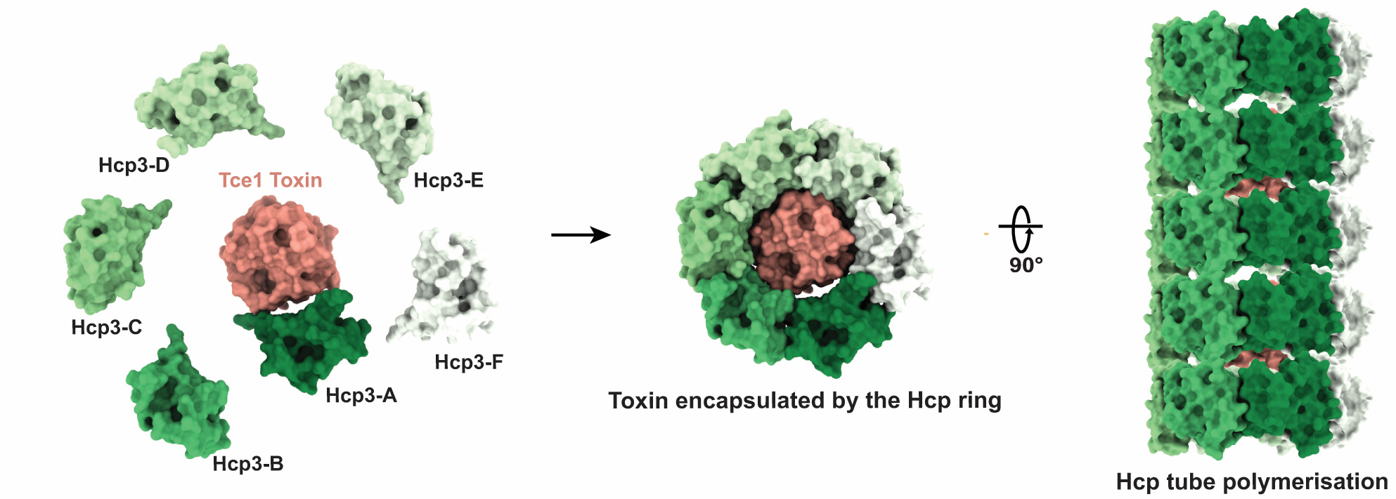
The Hcp proteins encapsulate the toxin and the complexes form a long tube which contracts to deliver the toxins. Credit: Imperial College London and NTU Singapore.
Using biochemical and cryo-electron microscopy analysis, they found that the T6SS is assembled in a step-by-step process. Toxins are first captured by a protein called Hcp. Five additional Hcp proteins then wrap around the toxin to form a ring. Each toxin-loaded ring resembles a flying saucer, with the toxin positioned in the centre. In some cases, two rings may be needed to fully enclose a larger toxin.
The loaded rings then stack on top of one another to form a long tube. When the system contracts, it propels the tube outwards like a harpoon, delivering the payload of toxins into a target cell.
The tube can carry different toxins, so a single firing event may deliver a cocktail of compounds.
“This bacterium does not just fire a single toxin. It loads a cocktail of toxins into a microscopic speargun and fires them in one strike, allowing it to attack different targets, including beneficial bacteria that normally live in the body, as well as the host’s own defence cells,” says co-corresponding author Prof Alain Filloux, research director of the Biofilms & Health Cluster at NTU’s Singapore Centre for Environmental Life Sciences Engineering, who is also from NTU’s SBS and LKCMedicine.
“This helps the bacterium colonise the host more effectively. If we can block this loading step in future, it could pave the way for approaches that disarm the bacterium and make it less able to cause disease.”
According to the researchers, harmless bacteria can also be engineered with a T6SS loaded with toxins to fight invading bacteria, such as antimicrobial resistant gut pathogens.
“What is exciting about this work is that we can now see, at near-atomic detail, how a bacterial toxin is physically captured and enclosed inside the building blocks of the T6SS,” adds Assoc Prof Tiago Dias da Costa from Imperial College London’s Department of Life Sciences at the Faculty of Natural Sciences, who co-led the study.
“High-resolution 3D images obtained from cryo-electron microscopy reveal that toxin loading is not a passive process, but a highly organised assembly pathway in which the secretion tube forms around its cargo. This gives us a molecular explanation for how P. aeruginosa prepares a cocktail of toxic effectors before firing them into competing microbes or host cells. By understanding this mechanism, we not only uncover a fundamental principle of bacterial cell biology but also identify new ways in which these systems might eventually be disrupted or repurposed.”
Watch a video on how P. aeruginosa loads and fires toxins:
Find more details in:
“The Mycobacterium abscessus cytochrome bcc:aa3 oxidase structure paves the way for an agent targeting subunit QcrB”, Nature Communications (2026), DOI: doi.org/10.1038/s41467-026-70805-5.
“Smooth-to-rough morphotype switching, a mechanism of phage resistance in Mycobacterium abscessus”, Proceedings of the National Academy of Sciences of the United States of America (2026), DOI: doi.org/10.1073/pnas.2531197123.
“Molecular basis of type VI secretion system effector loading,” Nature Microbiology (2026), DOI: doi.org/10.1038/s41564-026-02363-x.