
Mapping heart regeneration genes could pave the way for new treatments for heart disease
The findings could spark novel treatments that improve recovery after heart attacks.
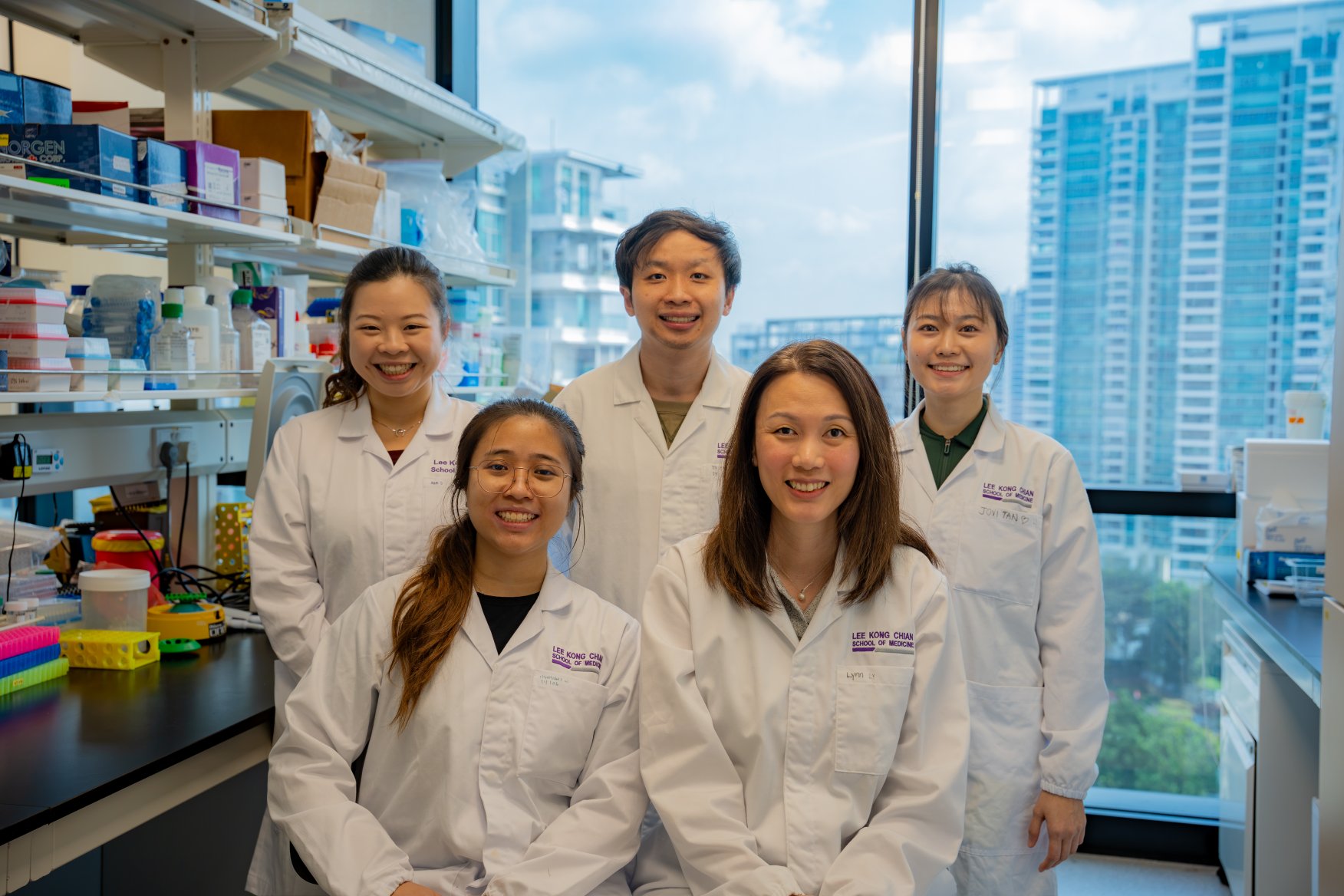 Front (from left): Christabel Chan, research assistant at LKCMedicine, and Asst Prof Lynn Yap. Back (from left): PhD students Samantha Lim, Leong Kye Siong and Jovi Tan. Credit: LKCMedicine.
Researchers led by Asst Prof Lynn Yap of the Lee Kong Chian School of Medicine at Nanyang Technological University, Singapore have uncovered new insights into how unspecialised cells, also known as stem cells, develop and mature into heart muscle cells. The findings could pave the way for new treatments for heart disease.
Published in Nature Cardiovascular Research, the study is one of the first to track the activity of genes at different points of the regeneration process.
The scientists also found that a protein called Midkine can repair damaged blood vessels and may help improve recovery after heart attacks.
Getting to the heart of the matter
Heart disease is one of the leading causes of death globally, accounting for a third of deaths worldwide[1]. In Singapore, about 22 people die from heart disease every day[2].
Heart disease occurs when blocked arteries prevent oxygen and nutrients carried by blood from reaching the heart muscle, which causes the cells to die. As heart muscle is unable to repair itself after a heart attack, the body produces scar tissue to patch the damage, like a band-aid. However, scar tissue has a limited ability to contract and can lead to heart failure over time.
Stem cells, the master builder cells of the body, can develop into any cell in the body and could potentially be used to repair damaged tissues. A type of stem cell called human pluripotent stem cell-derived cardiovascular progenitor cell (CVP) can develop into the different cells that make up the heart and could be used to regenerate heart muscle in patients with heart disease.
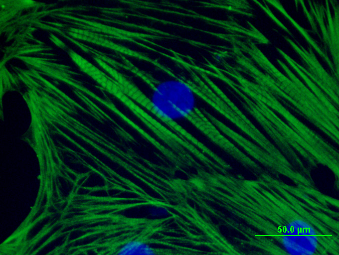
The scientists transplanted CVPs (above) into pig hearts and tracked the genes expressed by the cells as they matured into heart muscle cells. Credit: LKCMedicine.
To map this development, the researchers induced heart attacks in pigs and transplanted CVPs into the animals’ hearts. They then measured gene expression in the cells at various time points using a method called spatial transcriptomics.
Unlike conventional methods of measuring gene activity, the technique captures the activity of thousands of genes simultaneously while preserving information about where those genes are active within a tissue. In spatial transcriptomics, gene activity is determined at thousands of microscopic spots that capture RNA molecules produced when genes are switched on. Each spot acts like a tiny sensor to create a detailed map of gene activity.
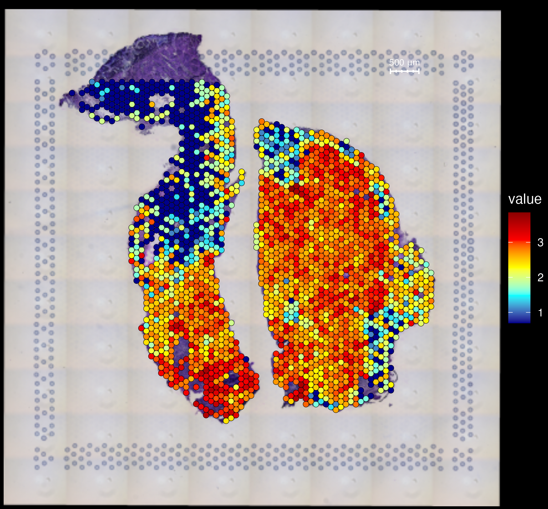
To detect gene activity after stem cell transplantation, a section of pig heart is placed on a slide containing probes that bind to RNA molecules produced by genes in the stem cells. In this molecular heat map, each coloured dot represents a small patch of tissue approximately 55 micrometres across. The colour indicates how actively the gene is being expressed – red dots signal high activity; blue dots signal low activity. Credit: LKCMedicine.
The researchers found that genes related to metabolism, energy generation, protein synthesis and heart muscle contraction were switched on after the cells were transplanted.
On the other hand, the activity of genes involved in scar tissue formation was reduced, suggesting that the stem cells successfully integrated into the damaged heart to improve tissue repair and reduce scarring.
Fixing a broken heart
The formation of blood vessels that supply the heart with oxygen and nutrients is vital for recovery after heart attacks. The researchers also identified a growth factor called Midkine produced by the transplanted stem cells that promotes the development of blood vessels.
First discovered in 1988, Midkine encourages cell growth and tissue repair. However, its role in recovery after heart disease is not well known.
In laboratory experiments, increasing the production of Midkine enhanced the ability of endothelial cells – the building blocks of blood vessels – to migrate and form new vessels.
Stem cells engineered to produce higher levels of Midkine also stimulated blood vessel formation in mice.
“Our study provides an unprecedented understanding of how stem cells interact with the damaged heart to regenerate heart muscle, which may accelerate the development of novel stem-cell-based therapies to treat heart disease,” said Asst Prof Yap.
“The findings also show that Midkine is crucial for healing after heart disease and resolves a decades-old debate on the role of Midkine in heart repair.”
“By identifying Midkine as a key protein that enhances blood vessel growth, this work suggests that targeting Midkine could offer a promising strategy to preserve heart function and potentially reduce the incidence and severity of heart failure," said Clinical Assistant Professor Julian Kenrick Loh, senior consultant with the Department of Cardiology and Director of the Coronary Care Unit at the National Heart Centre Singapore, who was not involved in the study.
The team has created a web application to make their gene expression data freely available and searchable (https://bit.ly/human-pig-mi-hearts-st). Their next focus is to further the understanding of how Midkine works, with the aim of harnessing it to treat heart disease.
Read more in “Spatiotemporal Transcriptomics of Human Cardiovascular Progenitors in Pig Hearts Identifies MIDKINE as a Positive Regulator of Neovascularization” in Nature Cardiovascular Research, DOI: doi.org/10.1038/s44161-026-00851-1.
All animal experiments were conducted with prior approval from SingHealth’s Institutional Animal Care and Use Committee (IACUC) and the ARRIVE Guidelines were followed.

.tmb-listing.jpg?sfvrsn=8d60272d_1)

.tmb-listing.jpg?sfvrsn=43b583b8_2)