
NTU scientists identify brain circuits that regulate impulsive behaviours
The findings could lead to more effective management of disorders such as attention-deficit hyperactivity disorder and addiction.
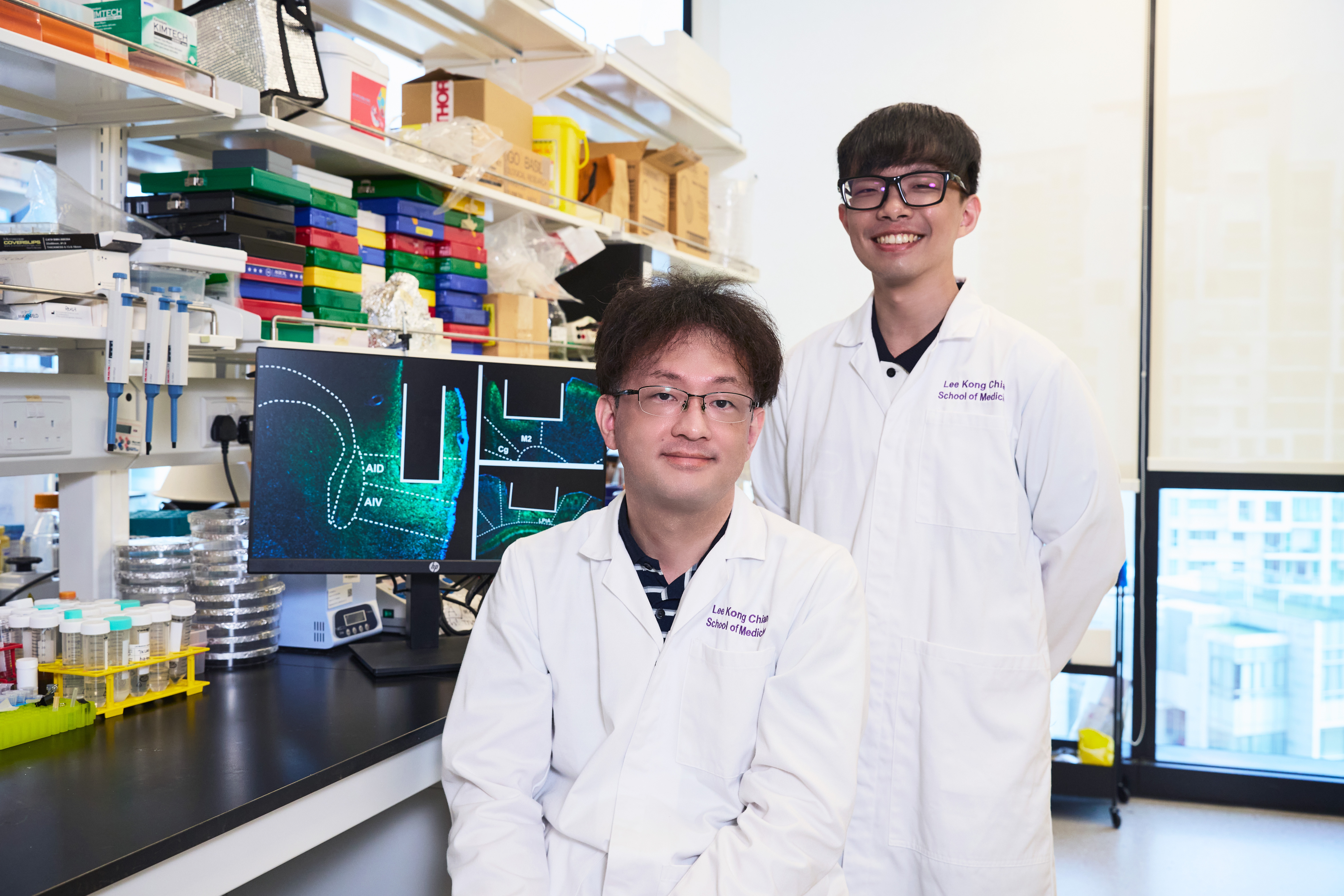
Scientists from the Lee Kong Chian School of Medicine at Nanyang Technological University, Singapore have uncovered how different brain regions work together to enable self-control – the ability to suppress impulsive behaviours and wait for the right moment to act. Their findings advance the understanding of conditions such as attention-deficit hyperactivity disorder (ADHD) and addiction, and could lead to more effective management of these disorders.
According to the researchers, this is the first time that this interplay underlying self-control has been found in the brain.
The findings were reported in Science Advances in October 2025.
Illuminating the secrets of self-control
Various neuropsychiatric conditions, such as impulse control disorders and ADHD, are associated with difficulties in impulse regulation. Individuals with such conditions may struggle with suppressing impulses and find it more difficult to pause and consider their actions before responding.
Three brain regions involved in reasoning and complex cognitive functions are thought to be crucial for our ability to control impulses – the anterior insular cortex (AIC), the posterior parietal cortex (PPC) and the dorsomedial frontal cortex (dmFC).
However, the exact mechanisms in this brain network that govern self-control are not well understood.
To examine self-control in mice, the researchers designed a task, in which the animals were trained to wait and withhold licking for brief seconds before receiving a liquid reward, mimicking the challenge of resisting an immediate impulse.
A sound was played to signal the start of the task, followed by a waiting period. An air-puff signalled the end of the waiting period, after which the mice could lick a water port to receive a water droplet as a reward. The task also included trials where no air-puff was delivered. In these instances, the mice had to rely on their internal timing to wait for a required duration before licking; if they licked too early, they would not receive a reward.
How long the mice successfully withheld the urge to lick served as a direct measure of their self-control.
The researchers then used optogenetics, a technique that uses light to control the activity of cells, to inactivate the brain regions involved in self-control and observed the impact of these areas on the impulsivity of the mice.
When the dmFC was inhibited, the mice waited for a shorter time before licking the water port, indicating that they were more impulsive. In contrast, when the AIC was inactivated, mice became more patient and waited longer before licking the water port.
Self-control also involves the ability to estimate and track the length of time that has passed, so that premature actions can be suppressed successfully. When the PPC was silenced, mice did not become more or less impulsive. Instead, it made their waiting behaviour less consistent, suggesting that the PPC supports self-control by stabilising timing rather than directly controlling impulsivity.
By imaging the calcium concentrations in neurons with fluorescent dyes, the researchers measured the activity of the neurons in the various brain regions. They discovered that some neurons in the PPC fired sequentially at specific elapsed times during the waiting period, much like the ticking of the clock as time progresses. These so-called “time cells” were specialised for tracking how much time had passed.
The researchers also examined the activity of neurons related to licking, a key measure of impulsiveness. Neurons that were less active during the licking period compared to the waiting period were classified as “motor-decreased”, or “motor-increased” if they were more active during the licking period.
They found that in the AIC, the suppressed activity of “motor-increased” neurons during the waiting period was associated with longer waiting times. On the other hand, the heightened activity of “motor-decreased” neurons in the dmFC was linked to more patient waiting.
These findings revealed that the three regions of the brain contribute to impulse control in distinct ways. The dmFC and AIC engage in a “push-pull” mechanism, where the dmFC promotes patience and the AIC regulates impulsivity, and their activity predicts how long the mice will wait. Meanwhile, the PPC plays a pivotal role in tracking how much time has elapsed.
“Our study provides one of the clearest demonstrations to date that different brain regions make complementary contributions to impulse control during waiting,” says Asst Prof Tsukasa Kamigaki, corresponding author of the study.
"By identifying the specific roles of different brain regions in regulating impulsivity, we are moving towards a more nuanced understanding of conditions such as ADHD, which could eventually lead to more targeted treatments of these disorders,” says psychiatrist and senior consultant Assoc Prof Jimmy Lee from the Institute of Mental Health in Singapore, who was not involved in the study.
“Importantly, this work reinforces that impulse control differences have clear neurobiological bases, which helps validate these as legitimate medical conditions rather than personal failings."
In the next step, the researchers aim to investigate which mechanisms are disrupted in various self-control related disorders, and to test treatments that target each of them.
Read more in “Dissociable cortical contributions to impulse control during waiting” in Science Advances, DOI: 10.1126/sciadv.adx4732.

.tmb-listing.jpg?sfvrsn=8d60272d_1)