Catch me if you can: how three infectious disease agents evade the immune system
NTU researchers are understanding how the respiratory syncytial virus, Chikungunya virus and malaria parasite escape being detected by the immune system.
COVID-19 cast a glaring spotlight on the devastating impact that infectious diseases can have on human health and society. At the height of the global pandemic, most of the world came to a standstill, and millions of lives were lost to the disease.
Although advances in health care have significantly reduced the global burden of infectious diseases, communicable diseases, like lower respiratory tract infections and malaria, remain among the top causes of premature death and ill health in developing countries. The growing threat of drug-resistant infections makes treating them more difficult, increasing the risk of disease spread and severe illness.
At the same time, changes in weather patterns caused by climate change are expected to lead to conditions that promote the breeding of insects that transmit infectious diseases, and their incidence will rise.
Amid the threat of Disease X, a severe global pandemic in the future that a yet unknown pathogen could cause, there is a need to develop more effective treatments for infectious diseases. Understanding how pathogens replicate, infect cells and evade the immune system may pave the way to treatments for both old and newly emerging infectious diseases.
Sticking around in airways
Like SARS-CoV-2, the virus that causes COVID-19, respiratory syncytial virus (RSV) infects the respiratory tract and causes symptoms that resemble the common cold. However, RSV can lead to more severe symptoms when it spreads to the lungs. In this context, RSV is the leading cause of viral pneumonia in young children, and the virus is responsible for several million hospitalisations and approximately 200,000 deaths in young children each year.
To better understand how RSV infects the respiratory tract, Assoc Prof Richard Sugrue from NTU’s School of Biological Sciences and his team have shed light on how a little-understood protein called small hydrophobic (SH) protein facilitates virus infection.
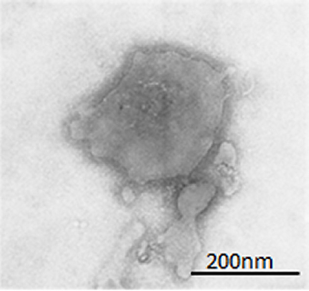
Transmission electron microscope image of an RSV particle. Image credit: Assoc Prof Richard Sugrue.
Studying human nasal epithelial airway cells infected with RSV, the researchers found that the SH protein is incorporated into new virus particles produced by the infected cells, supporting the proposed role of the protein in evading the cell’s early immune responses.
In later stages of the infection, the SH protein also causes ciliated epithelial cells to lose cilia — microscopic hair-like structures extending from each cell’s surface. In the respiratory tract, cilia are an essential part of the self-clearing mechanism of the human respiratory tract that sweeps inhaled microorganisms and debris up and out of the airways.
The researchers predict that the loss of cilia helps RSV colonise the upper airway by reducing the ability of the host to clear the virus from the upper airways.
“To date, antibodies are the most effective treatment option to protect children against RSV-related complications, so our investigation may open the door to new therapies that target the SH protein to treat the infection,” Sugrue said.
The study was published in the journal Virology in January 2023.
Replicating undetected
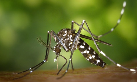
As the globe warms due to climate change, higher temperatures will increase mosquito populations, potentially causing a rise in transmission of mosquito-borne infectious diseases, such as dengue, Zika, Chikungunya and malaria.
A viral infection spread by Aedes mosquitoes, Chikungunya, affects several organs. Complications of the disease include fever, fatigue, muscle and joint pains.
In infected cells, the genetic material of the Chikungunya virus, ribonucleic acid (RNA), is duplicated inside a compartment called the replication complex (RC). The RC protects the newly synthesised RNA from being detected and destroyed by the host immune system.
Four nonstructural proteins (nsPs) — nsP1, nsP2, nsP3 and nsP4 — produced by the Chikungunya virus assemble to form the RC. However, researchers know little about its molecular architecture and how it works.
Using cryo-election microscopy to visualise the RC, NTU scientists discovered that it was a circular structure resembling a flying saucer.
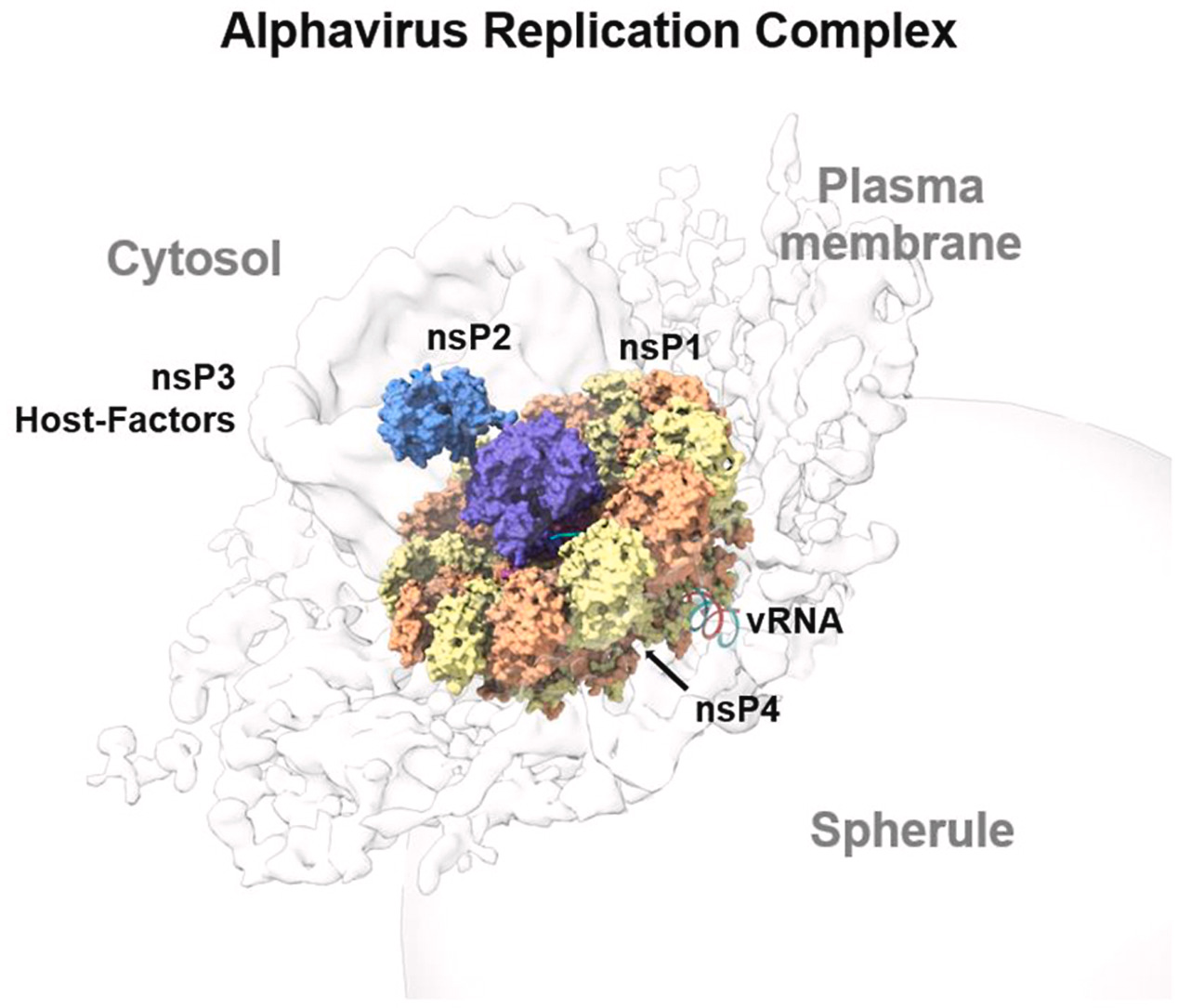
Four nonstructural proteins make up the Chikungunya replication complex. Image credit: Assoc Prof Luo Dahai.
The researchers demonstrated that nsP1 promoted the RNA polymerase activity of nsP4 — required for synthesising RNA during virus replication. The binding of nsP2 to the nsP1 + nsP4 complex further stabilises the complex and boosts its activity, catalysing the chemical reactions essential for RNA synthesis.
On the other hand, nsP3 is likely essential for forming spherules, which are dedicated membrane-like structures for virus replication formed on the surface of infected cells.
“Our findings provide an unprecedented understanding of the virus replication strategy in general, which may be applied to developing new antiviral drugs for virus infections such as dengue and COVID-19,” said Assoc Prof Luo Dahai, who led the study published in Science Advances in November 2022.
Escaping white blood cells
Malaria, caused by the Plasmodium parasite, is another infection spread by mosquitoes that is a public health concern, especially in developing regions where the disease is endemic. The disease has plagued humans for thousands of years, with the earliest reports of the infection dating back more than 2000 years.
The malaria parasite’s ability to evade the host immune system makes developing vaccines for the disease particularly challenging. Furthermore, the effective treatment of malaria is hampered by its increasing resistance to currently available antimalarial drugs, contributing to its spread.
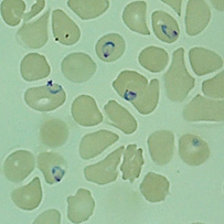
Red blood cells infected with malaria parasites (blue rings). Image credit: Prof Peter Preiser.
In a study published in Nature Communications in July 2022, NTU scientists uncovered how some proteins expressed by the parasite protect it from being cleared by white blood cells. The study in mice extends existing knowledge of the parasite from in vitro studies using cell cultures.
The scientists infected mice with Plasmodium falciparum, the deadliest species of the parasite that causes malaria in humans, and found that it produced proteins that helped it survive inside the host.
A protein produced by the parasite on its surface, called var2csa, helps it avoid being killed by white blood cells known as natural killer cells and macrophages.
The researchers suggest that var2csa helps the parasite fly under the immune system’s radar as it does not activate the immune responses required to eliminate the parasite. As a result, white blood cells are less able to get rid of it.
“With new evidence of how the malaria parasite escapes the immune system of the host, targeting the proteins crucial for immune evasion may offer a unique approach to treating malaria,” said Prof Peter Preiser of NTU’s School of Biological Sciences, who led the study.
Read the story on Lab+Life Scientist.
-and-dr-dorrain-low-from-ntu-singapore's-lkcmedicine.tmb-listing.jpg?Culture=en&sfvrsn=b389b500_1)
.tmb-listing.jpg?Culture=en&sfvrsn=82921582_1)

.tmb-listing.jpg?Culture=en&sfvrsn=ba129532_1)