Soft electrode array maps electrical signals travelling through plants
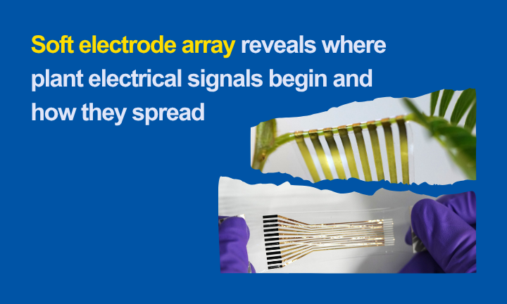
A low-drift, conformal electrode array, developed by NTU and A*STAR researchers, reveals where plant electrical signals begin, how they travel and how they relate to visible responses.
Collaborative feature with Prof Xiaodong Chen, and Dr Li Wenlong, A*STAR IMRE and NTU School of Materials Science and Engineering
Published in Nature Electronics May 2026
Touch the leaves of Mimosa pudica and the plant folds within seconds. Behind that visible movement is an electrical signal travelling through the plant - but conventional electrodes usually record at only one or two points. They can show that an electrical event occurred without revealing, in detail, where it began, which direction it travelled or how its passage related to the plant's response.
Researchers from NTU's School of Materials Science and Engineering and their collaborators have developed a soft electrode array that addresses this measurement gap. Wrapped around a plant stalk, the array records electrical activity at multiple positions. A bioelectronic model and interpolation method then turn these surface-potential measurements into colour-coded maps that can be overlaid on a video of the plant.
The result is a moving picture of electrical activity across space and time. Rather than listening for a plant signal at a single location, researchers can follow the equivalent of a stadium wave: where it starts, how it moves and what happens when it encounters damage or another signal.
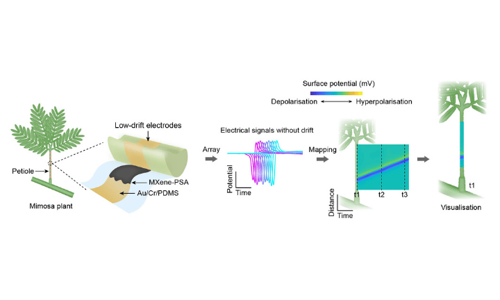 |
| How the low-drift electrode array wrap on a Mimosa petiole records surface potential signals without drift. |
Solving a difficult plant interface
Reliable mapping first required the team to solve a materials problem. Mimosa petioles are slender and highly curved, with a curvature of approximately 3 mm⁻¹. A high-density array must wrap securely around this surface without becoming so stiff or heavy that it interferes with the plant's natural movement.
Wet gels and hydrogels can improve electrical contact, but dehydration can undermine long-duration measurements and dense patterning. Dry electrodes are easier to pattern, yet insufficient contact with curved plant surfaces can introduce baseline drift and movement artefacts. Across several channels, these instabilities can distort the reconstructed signal map.
The researchers created a dry composite from Ti3C2Tx MXene, a conductive two-dimensional material, and a pressure-sensitive adhesive. Printed onto a thin gold, chromium and polydimethylsiloxane structure, the composite combines conductivity with the adhesion and flexibility needed to form a full 360-degree wrap around the Mimosa petiole. The complete device weighs less than 60 mg, and the electrode array remained intact when stretched to 100 per cent strain.
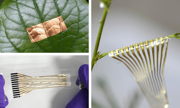 |
| Dry composite from Ti3C2Tx MXene on a leaf and photographs of the low-drift electrode array |
On an undisturbed Mimosa, the MXene-adhesive electrodes recorded a drift of 77.5 ± 11.8 microvolts per minute. This was approximately half the drift measured with conventional gold-polyacrylamide electrodes and one-sixth that of silver/silver-chloride gel electrodes. The system also recorded continuously for 15 hours. Separately, the composite film retained useful electrical conductivity after four months of ambient storage.
Turning measurements into moving maps
Stable contact alone was not enough. Signals measured at a plant's surface depend on electrode position and spacing, as well as the electrical activity within the tissue. The NTU team therefore developed a bioelectronic model to explain how different electrode configurations capture a propagating action potential and to identify the most suitable arrangements for measuring velocity, amplitude or spatial detail.
Using multichannel measurements and a customised interpolation algorithm, the researchers reconstructed spatiotemporal maps of electrical activity along the petiole. In experiments where the plant was stimulated at the apex, middle, base or both ends, the maps revealed signals travelling forward, backward or in both directions. When signals travelling from opposite directions met, they annihilated on collision, consistent with the refractory period associated with action potentials.
The mapping also separated a Mimosa response into three stages: sensing the stimulus, transmitting an electrical signal and initiating visible movement. When the plant was stimulated, the petiole drooped only after the electrical signal reached its base. Across touch, cold and heat experiments, the sensing and signalling stages lasted longer than the subsequent physical response, which typically occurred in less than half a second.
Different stimuli, different observations
The platform allowed the team to quantify how Mimosa responded under the reported experimental conditions. A mechanical force of approximately 200 millinewtons corresponded to a 50 per cent probability of triggering an action potential. The experiments also identified response thresholds of about 5 degrees Celsius for cold-water stimulation and 75 degrees Celsius for hot-water stimulation. These are controlled laboratory thresholds and should not be interpreted as general crop-stress limits in natural environments.
| Visualisation of real-time electrical signal Vsp on Mimosa petiole under different mechanical and temperature stimuli. | |
Changes in light produced a different spatial pattern. Within seven seconds of switching off intense light, a localised electrical signal emerged near the base of the petiole; within ten seconds, the petiole drooped. The observation suggests that local electrical activity may contribute to Mimosa's response to transitions between light and darkness, although the underlying mechanism remains to be clarified.
The researchers also partially cut a Mimosa petiole and followed its response over time. Immediately after the injury, the signal weakened near the damaged region and did not pass through it, leaving the petiole upright. Electrical transmission and the drooping response returned after 15 hours, although the signal moved more slowly past the injured site. This allowed the team to distinguish phases of impairment, recovery and subsequent fatigue.
| Visualisation of real-time electrical signal Vsp on Mimosa petiole under high intense light stimulation and before and after a partial cut. | |
Extending the view beyond Mimosa
To examine whether the approach could reveal electrical activity in other plants, the researchers mapped wound-induced signals in Arabidopsis thaliana mutants and in choy sum. In Arabidopsis, an eight-channel configuration resolved two components of the wound response and showed how mutations affecting particular ion channels altered their propagation. The glutamate receptor double mutant glr3.3 glr3.6 lacked detectable travelling depolarisation waves, supporting the essential role of these channels in long-distance electrical signalling.
In choy sum, a 16-channel array revealed two distinct wound-response modes across 28 measurements. In one, a well-defined depolarisation wave travelled along the full petiole. In the other, a slower and broader wave weakened before reaching the base. The two modes appeared at similar frequencies, but the biological factors determining them remain unknown.
These experiments demonstrate the platform's value as a research tool rather than a completed agricultural product. By moving from isolated readings towards dynamic maps, it gives plant scientists a way to ask more precise questions about signalling, injury, recovery and the roles of individual ion channels.
Towards more complete maps of plant health
For plant biologists and crop-breeding researchers, the platform opens a new window into how plants process and transmit information. Researchers can examine not only whether a plant responds to a stimulus, but where its electrical response begins, how it travels and how signalling changes following injury or genetic modification. Extended across more plant varieties and growing conditions, this capability could add an electrical dimension to phenotyping—complementing visual and physiological measurements when comparing responses to environmental stress.
The work also creates opportunities for collaboration with companies developing plant-research instruments, flexible and printed electronics, low-noise bioinstrumentation and advanced electrode materials. Its low-drift, conformal electrode array provides a foundation for future portable and wireless plant-monitoring technologies.
The next opportunity is to extend the platform beyond the linear, one-dimensional plant structures studied in the laboratory. Leaves and other organs have more complex two- and three-dimensional geometries, requiring electrode systems that can conform to a wider range of surfaces. Wireless read-out, portable electronics and longer-term plant compatibility will also be needed before the technology can be evaluated under natural field conditions.
Addressing these challenges could ultimately extend spatial electrical mapping into crop phenotyping, seed development, crop protection, greenhouse cultivation, vertical farming and precision agriculture. By complementing cameras and environmental sensors with information about a plant’s internal response, future systems could give researchers and growers a more complete picture of how plants respond to their environment.
More about the publication
Published in Nature Electronics, the study brought together researchers from NTU's School of Materials Science and Engineering, School of Biological Sciences, School of Mechanical and Aerospace Engineering, School of Electrical and Electronic Engineering, and Institute for Digital Molecular Analytics and Science, as well as A*STAR's Institute of Materials Research and Engineering.
The research was supported by the A*STAR MTC Programmatic Funding Scheme and the National Research Foundation (NRF), Prime Minister's Office, Singapore, through the Singapore Hybrid-Integrated Next-Generation μ-Electronics (SHINE) Centre and an NRF Investigatorship.
Paper: Direct visualisation of environment-stimulated electrical signals in plants using low-drift dry electrodes
https://doi.org/10.1038/s41928-026-01642-z
More about Prof Chen Xiaodong
 | Distinguished University Professor, NTU Singapore
School of Materials Science and Engineering Plant bioelectronics | Flexible and conformal devices | Artificial senses | Bioelectronic interfaces | MXene-based conductive composites |