Stem Cell Discovery Brings Hope for Novel Treatment for Heart Disease
Text: Camellia Yong | Photos: Aaron Koo
Constructing a sturdy house requires more than laying bricks. Cement binds the structure together, while unobstructed pipes ensure water reaches every corner of the home. In much the same way, recovery after a heart attack depends not only on repairing damaged heart muscle but also on healing the surrounding tissue and restoring blood flow through affected blood vessels.
Our researchers at NTU’s Lee Kong Chian School of Medicine are now exploring how stem cells could help achieve both.
What happens after a heart attack?
After a heart attack, part of the heart muscle is deprived of oxygen because blood flow is blocked. As heart muscle is unable to repair itself, the body produces scar tissue to patch the damaged tissue. However, scar tissue has a limited ability to contract and can lead to heart failure over time. 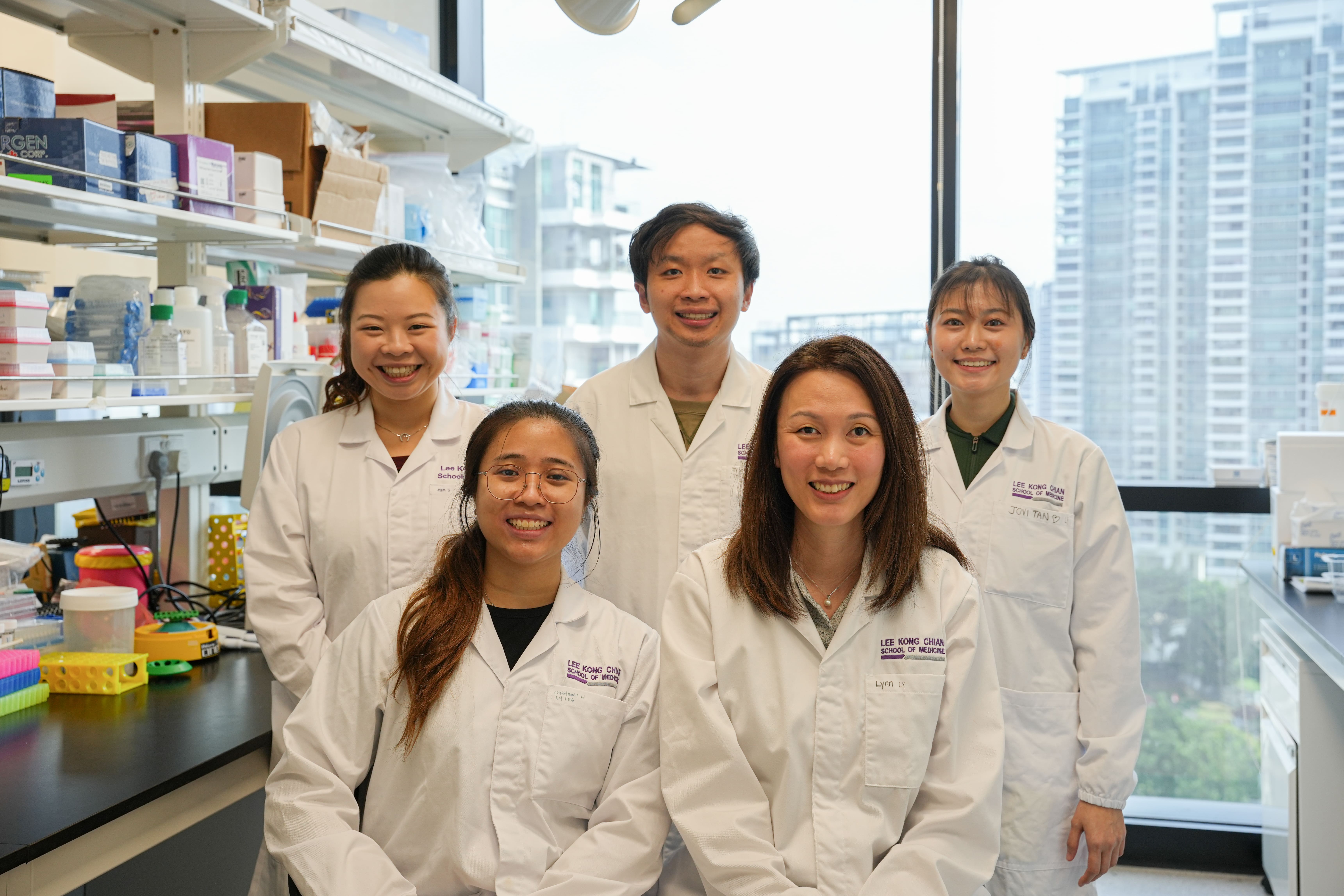 Front (from left): Christabel Chan, research assistant at LKCMedicine, and Asst Prof Lynn Yap. Back (from left): PhD students Samantha Lim, Kye Siong Leong and Jovi Tan. Credit: LKCMedicine.
Researchers led by Assistant Professor Lynn Yap discovered that human cardiac precursor cells (known as cardiovascular progenitor cell or CVP) can develop into the different cells that make up the heart and regenerate heart muscle in patients with heart disease.
They also found that a protein called Midkine was secreted from the injected CVP that can help repair damaged blood vessels and enhance the recovery after heart attacks. The study, published in Nature Cardiovascular Research, is one of the first to track the activity of genes at different points of the regeneration process.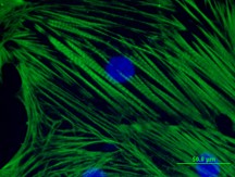 The scientists transplanted CVPs into pig hearts and tracked the genes expressed by the cells as they matured into heart muscle cells.
Mapping the discovery
To validate and visualise the study, the researchers transplanted CVPs into pig hearts damaged by a heart attack. They then measured the activity of the genes in the cells at various time points using a method called spatial transcriptomics.
The researchers found that genes related to metabolism, energy generation, protein synthesis and heart muscle contraction were switched on after the cells were transplanted. On the other hand, the activity of genes involved in scar tissue formation was reduced, suggesting that the stem cells successfully integrated into the damaged heart to improve tissue repair and reduce scarring.
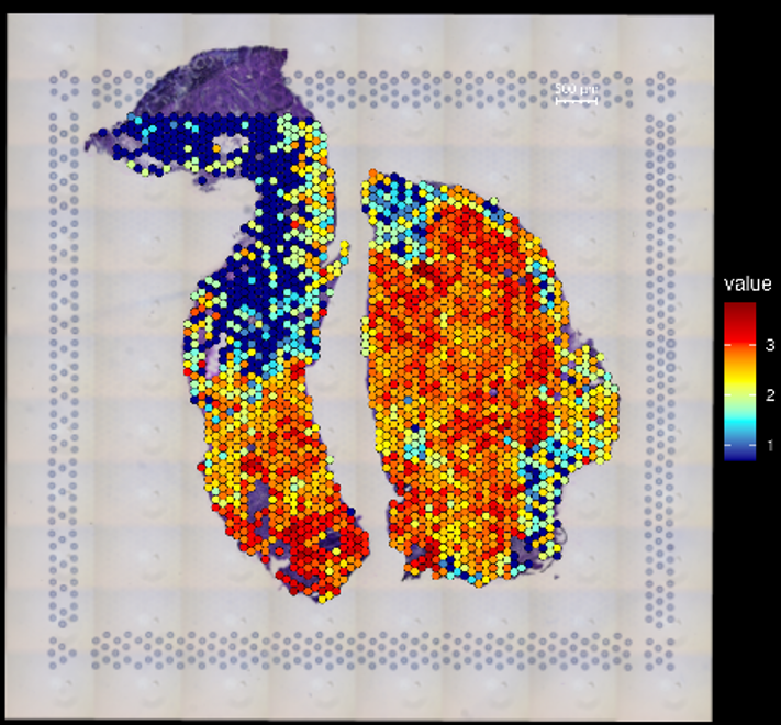 The spatial transcriptomics shows the gene activity after stem cell transplantation. The colour indicates how actively the gene is being expressed – red dots signal high activity in the recovery of heart muscle; blue dots signal low activity in the formation of scar tissue.
“Our study provides an unprecedented understanding of how stem cells interact with the damaged heart to regenerate heart muscle, which may accelerate the development of novel stem-cell based therapies to treat heart disease,” said Asst Prof Yap.
“The findings also show that Midkine is crucial for healing after heart disease and resolves a decades-old debate on the role of Midkine in heart repair.”
What’s next?
Asst Prof Yap and her team are presenting this research at international conferences such as Stem Cell and Development Biology Symposium organised by International Society for Stem Cell Research (ISSCR) and Allen institute in Seattle, USA and Basic Cardiovascular Sciences (BCVS) organised by American Heart Association (AHA) in Boston, USA.
Next, they will focus on furthering their understanding of how Midkine works, with the aim of harnessing it to treat heart disease.
Read more in “Spatiotemporal Transcriptomics of Human Cardiovascular Progenitors in Pig Hearts Identifies MIDKINE as a Positive Regulator of Neovascularization” in Nature Cardiovascular Research, DOI: doi.org/10.1038/s44161-026-00851-1.
All animal experiments were conducted with prior approval from SingHealth’s Institutional Animal Care and Use Committee (IACUC) and the ARRIVE Guidelines were followed.

.tmb-listing.jpg?sfvrsn=40968450_1)