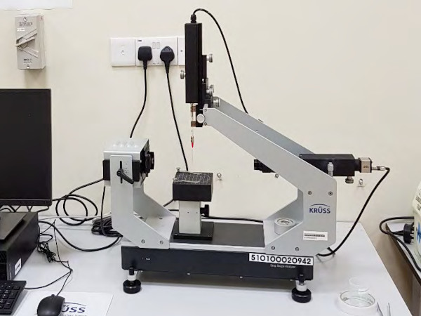
Atomic and Nanoscale Imaging/Microscopy
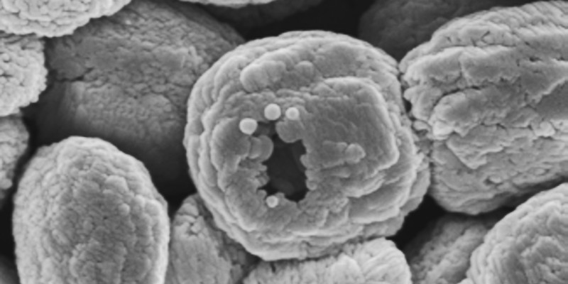
The following instruments are housed in N1.2 and N1.3.
Contact Information
Usage of the facility is handled through the Central Equipment Booking System.
For inquiries about specific instruments, please contact the following staff:
| JSM 6701F SEM / JSM 6390LA SEM / ICON2-SYS AFM | Dr WANG Xiujuan Phone: (65) 6790-6495 Email: [email protected] |
| JEM 3010 TEM / JEM 2100PLUS TEM | Dr LUA Shun Kuang / Dr WANG Xiujuan Phone: (65) 6316-8814 / 6790-6495 Email: [email protected] / [email protected] |
| JSM6700F SEM/ Zeiss LSM 800 CM | Dr YU Shucong Phone: (65) 6790 4064 Email: [email protected] |
| DSA25 Contact Angle Analyzer | Mr QUEK Kiat Yong, Jason Phone: (65) 6513-7684 Email: [email protected] |
Equipment List
JEOL JSM 6700F/6701F
- High resolution of secondary electron image (SEI and LEI).
- High magnification x100 (WD 25 mm) to x650,000 (WD 8 mm)
- Accelerating voltage 0.5 to 30 kV
- Electron gun field emission with cold cathode
- Emission current 2, 5, 10, 20 µA; usually 10 µA
- High stability large eucentric specimen stage with motorized control
- X-axis 70 mm; Y-axis 50 mm; Z-axis 25 mm; Rotation 360° ; Tilt -5° to +60°
- Liquid N2 cold trap
- Oxford X-Max Energy Dispersive X-ray Analyser (EDS) offering seamless observation and micro-analysis
Location: N1.3-B3-19a (6700F), N1.2.B5-10 (6701F)

JEOL JSM 6390LA
- Magnification x5 (WD 48 mm) to x300,000 (WD 8 mm)
- Automatically corrected for accelerating voltage and WD changes
- Probe current 1 pA to 1 uA
- Fully automatic system control
- Ultimate pressure in gun chamber 0.1 mPa order at HV mode; 1 mPa order at LV mode
- X-axis 80 mm; Y-axis 40 mm; Z-axis 48 mm; Rotation 360° ; Tilt -10° to +80°
Location: N1.2-B5-10
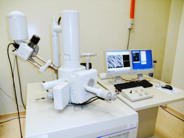
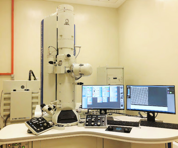
JEOL JEM 3010 TEM
- Accelerating Voltage 300 kV
- Magnification 4000x to 1200000x
- Electron gun LaB6 filament
- Condenser aperture, objective aperture and field limiting aperture
- OL polepiece HRP
- Polepiece lattice resolution 0.143 nm; point resolution 0.19 nm
- 5 spot sizes and 3 α angles
- Gatan Orius832 CCD camera
Location: N1.2-B5-10
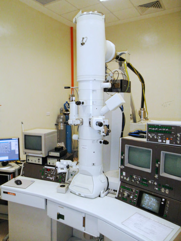
JEOL JEM 2100PLUS TEM
- Accelerating Voltage 200 kV
- Magnification 30x to 1500000x
- Electron gun LaB6 filament
- Condenser aperture, objective aperture and field limiting aperture
- Polepiece lattice resolution 0.14 nm; point resolution 0.23 nm
- 5 spot sizes and 3 α angles
- BF and DF STEM
- Gatan RIO camera
Location: N1.2-B5-10
Bruker ICON2-SYS AFM
- Anti-vibration hood
- Operating modes: Contact mode, Tapping mode and Force mode
- Scan Axes: X&Y 100 μm; Z >10 μm sensored travel
Location: N1.2-B5-10
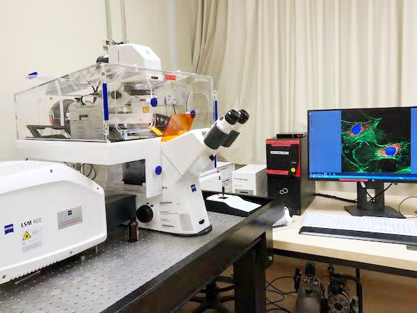
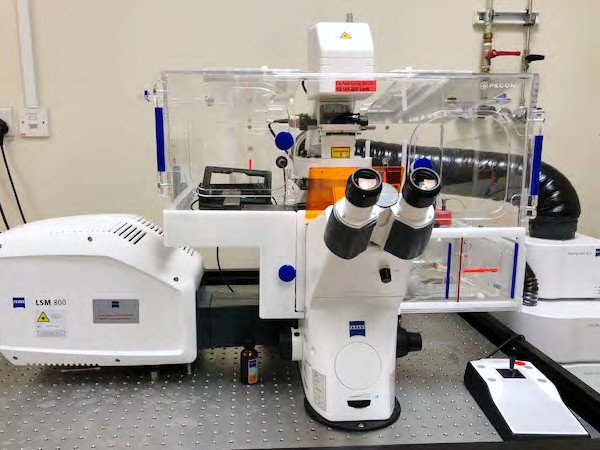
Zeiss LSM 800 Confocal Microscope
- Four Diode Lasers at 405/488/561/640 nm
- Two high sensitivity Gallium arsenide phosphide (GaAsP) detectors and one Airyscan detector for
super-resolution - Airyscan detector increases lateral resolution of an image by 1.4x without software post-processing, and increases sensitivity of detection
- Scanning speed: Up to 8 fps with 512x512 pixels
- Objectives: 5X, 10X, 20X, 40X, 63X, 100X
- Motorised stage with Z section scanning (100-200μm), Z1 has a 10nm step size with +/- 10 nm repeatability
- Live-cell stage equipment with incubation chambers and good environmental control
- Fluorescence Energy Transfer (FRET), Recovery After Photo bleaching (FRAP)
- Stitching ability for photo montages
Location: N1.3-B3-19b
KRUSS DSA25 Contact Angle Analyzer
- Contact angle measurements
- Surface and interfacial tension measurements
- Wettability and absorption measurements
- Computer-controlled syringe pump
- Computer-controlled lighting
- Adjustable specimen stage
Location: N1.2-B5-10
