
Microscopy Facility

The SBS Microscopy Facility is a shared research platform providing advanced microscopy technologies and expert support for life science research. Our services include instrument access, hands-on training, assisted imaging, image acquisition and analysis, and consultation on experimental design and imaging workflows.
Our facility is open to all NTU faculty, staff, students, and external collaborators.
For enquiries about imaging services, research collaborations, instrument training, or facility charges, please contact us.
CONTACT INFORMATION
| Location of Facility | School of Biological Sciences |
| Contact Person | Dr. Lai Soak Kuan |
AVAILABLE EQUIPMENT
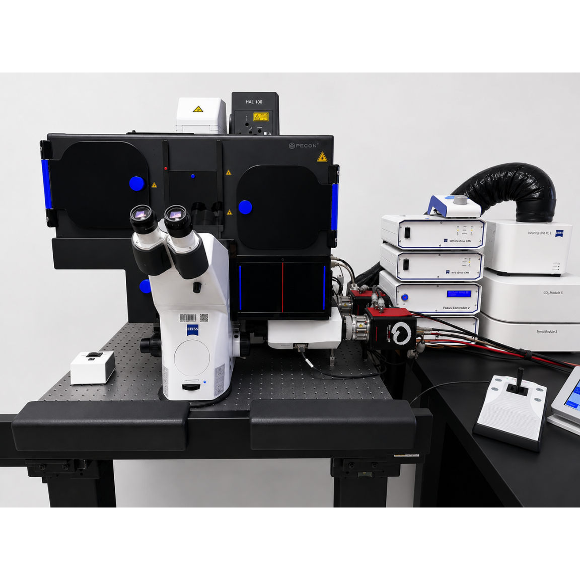
Elyra 7
Advanced super-resolution imaging platform offering Lattice SIM for fast, gentle imaging and SMLM for nanoscale molecular localization.
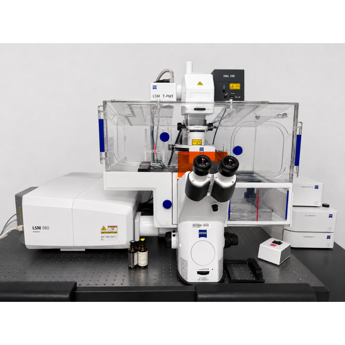
LSM 980 with Airyscan 2 Confocal
High-performance confocal microscope with Airyscan 2 for sensitive, high-resolution 3D and super-resolution fluorescence imaging.
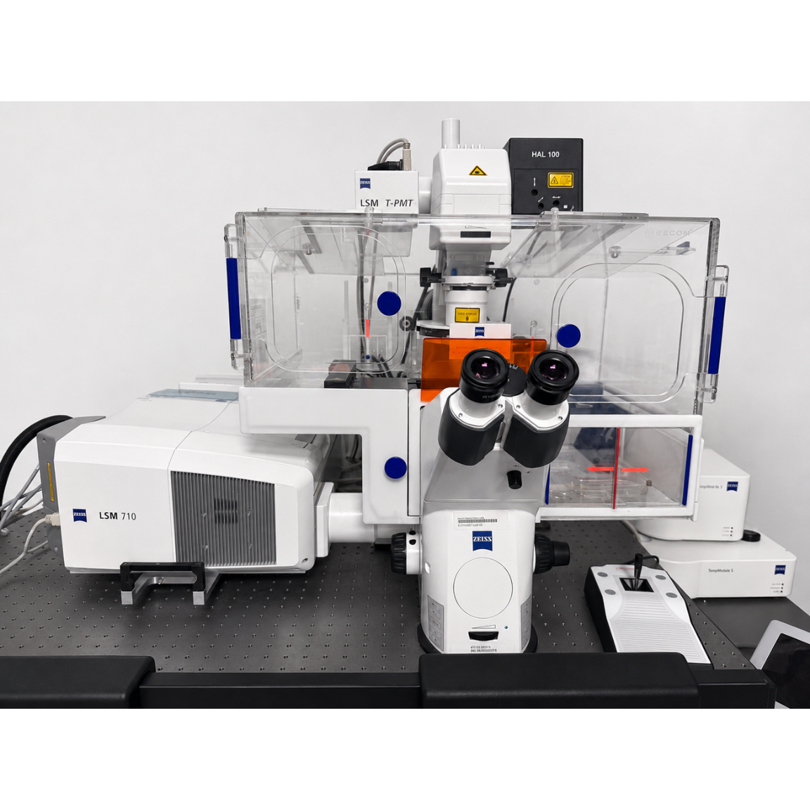
LSM 710 Confocal
Versatile confocal microscope for high-quality fluorescence imaging and optical sectioning of fixed and live biological samples.
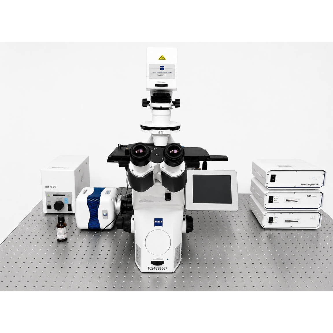
Laser TIRF3
Total internal reflection fluorescence (TIRF) microscope for highly sensitive imaging of molecular and cellular events occurring at or near the cell membrane.
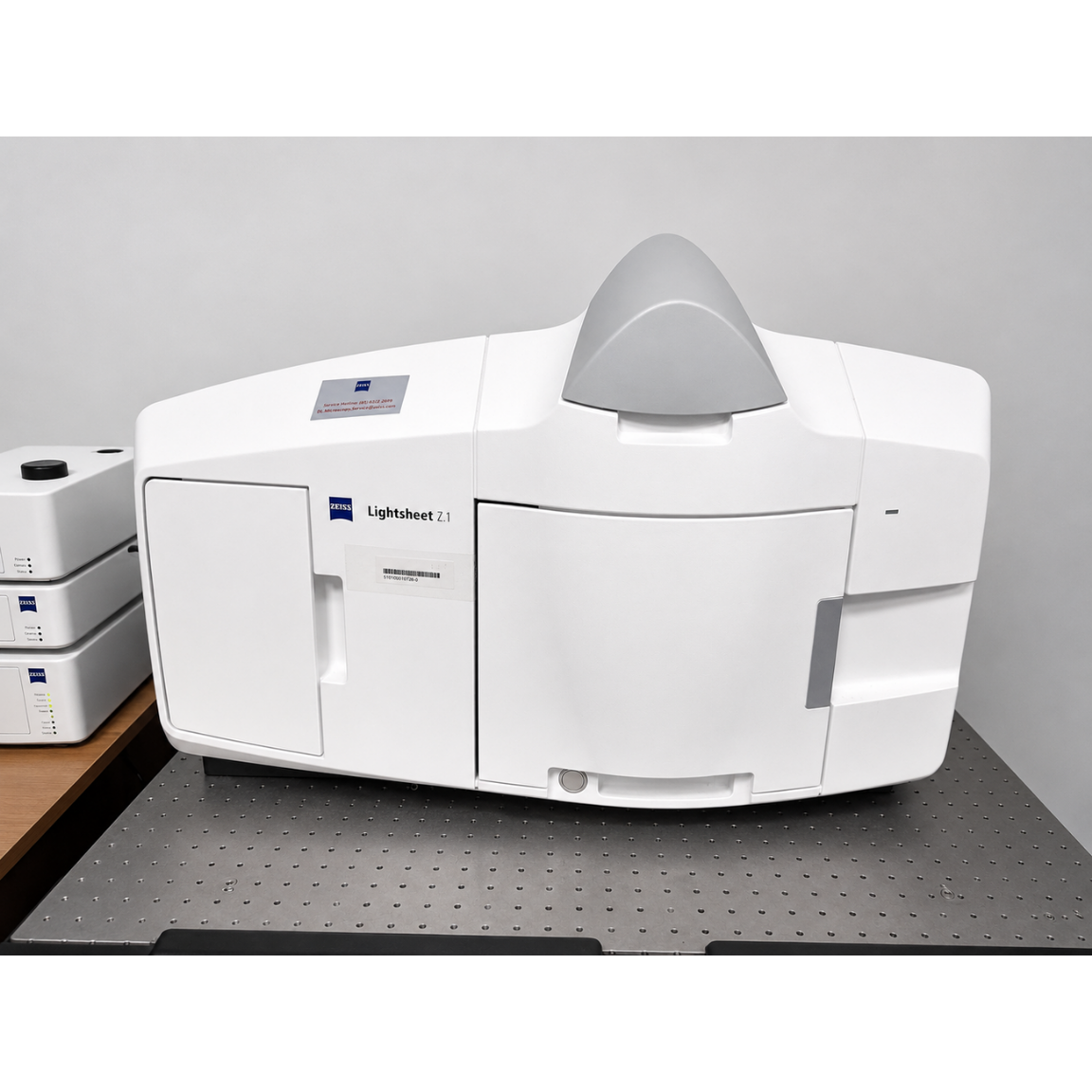
Lightsheet Z.1
Gentle and rapid 3D fluorescence imaging of large biological specimens with reduced phototoxicity and photobleaching.
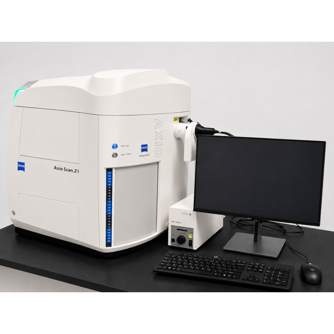
Axioscan Z.1
Automated scanning of multiple microscope slides for fluorescence and brightfield whole-slide imaging.
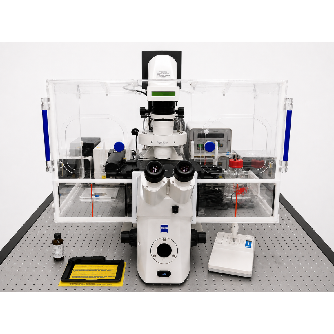
Live Cell Observer I
Widefield fluorescence microscope for routine imaging.
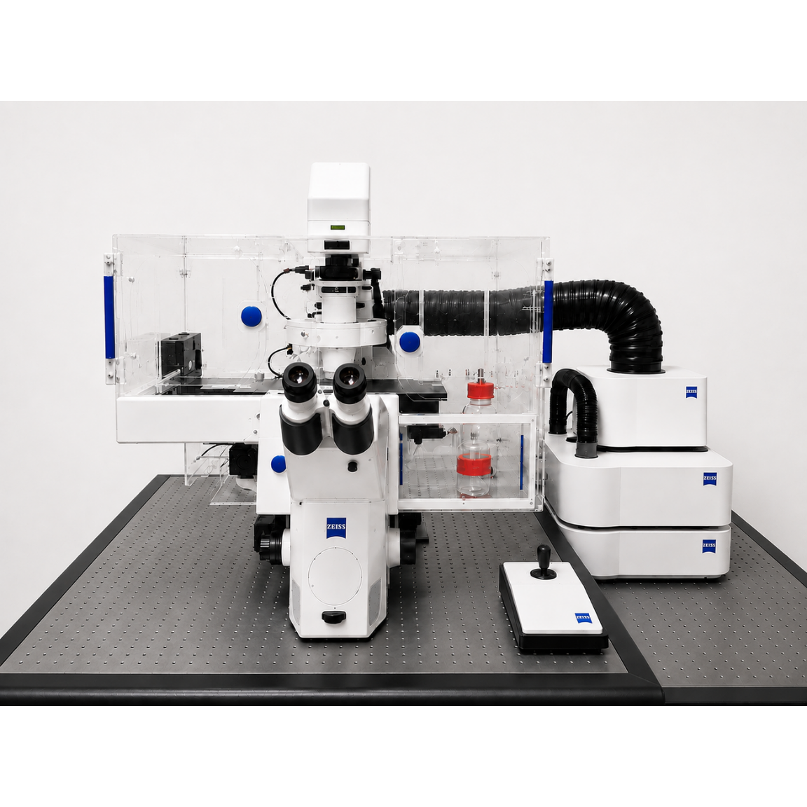
Live Cell Observer II
Widefield fluorescence imaging system with environmental control for time-lapse and live-cell imaging experiments.
SteREO Lumar.V12
Low-magnification fluorescence stereomicroscope for imaging and manipulation of embryos, tissues, and small model organisms.
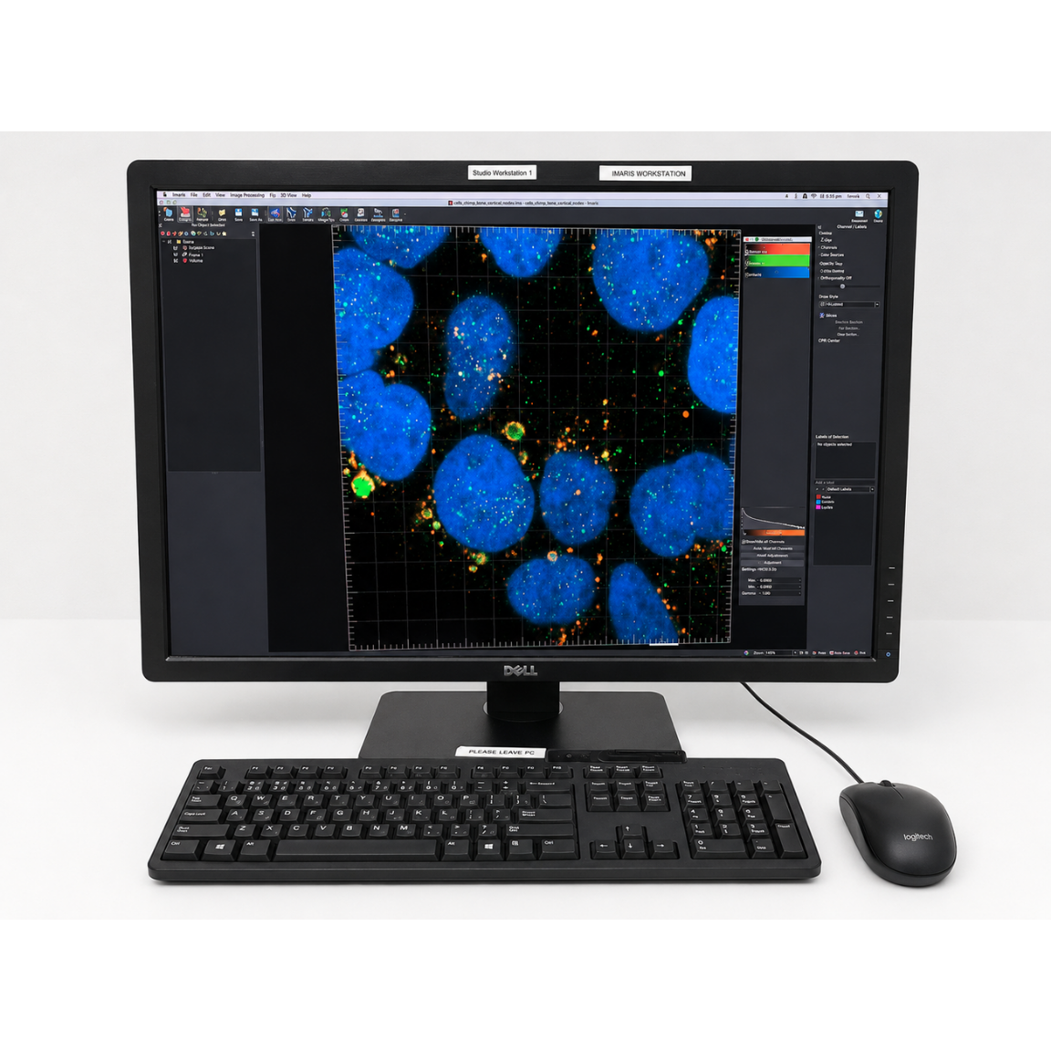
IMARIS
Advanced software for 3D/4D visualization, segmentation, tracking and quantitative analysis of microscopy datasets.
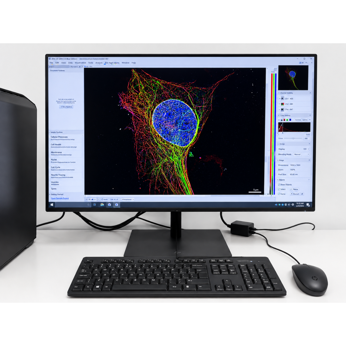
ARIVIS PRO
Powerful image-analysis platform for visualization, segmentation and quantitative analysis of large multidimensional microscopy datasets.