Microscopy
It is open to all NTU faculty, staff and students as well as external users.
CONTACT INFORMATION
| Location of Facility
| School of Biological Sciences 02S-071 |
| Contact Details
(For discussion on services and collaborations) | (Dr) Lai Soak Kuan
Email Address: [email protected] (for charges and enquiries) |
TYPES OF MICROSCOPES AVAILABLE

SPECIFICATIONS
Microscope Body
- Axio Observer.Z1
- Environmental Chamber for temperature control
Objectives
- EC Plan-NeoFluar 10x/0.30
- EC Plan-NeoFluar 20x/0.50
- EC Plan-NeoFluar 40x/1.30 Oil DIC
- Plan-Apochromat 63x/1.40 Oil DIC
- EC Plan-NeoFluar 100x/1.3 Oil
Light Source
- Halogen lamp for transmitted light
- X-Cite 120 illuminator for reflected light
- Argon (458,488,514nm)
- HeNe 633nm
- DPSS 561nm
- Diode 405nm
Filter Sets
| Filter set | Ex | Em |
| Zeiss Filter set 38 (GFP) | BP 470/40 | BP 525/50 |
| Zeiss Filter set 43 (dsRed) | BP 550/25 | BP 605/70 |
| Zeiss Filter set 49 (DAPI) | G365nm | BP 445/50 |
Detectors
- 2 standard PMTs
- Airyscan detector
Software
- Zen 2.1 (black)

SPECIFICATIONS
>Microscope Body
- Axiovert 200M
Objectives
- Plan-NeoFluar 10x/0.30
- Plan-Apochromat 20x/0.6 Ph2
- LD Achroplan 40x/0.6 corr Ph2
- Plan-Apochromat 63x/1.40 Oil DIC
- Plan-NeoFluar 100x/1.3 Oil Ph3
Light Source
- Halogen lamp for transmitted light
- HBO100 for reflected light
- Argon (458,488,514nm)
- HeNe 543nm
- HeNe 633nm
- Diode 405nm
Filter Sets
| Filter set | Ex | Em |
| Zeiss Filter set 01 (Blue) | BP 365/12 | LP 397 |
| Zeiss Filter set 09 (Green) | BP 450-490 | LP 515 |
| Zeiss Filter set 15 (Red) | BP 546/12 | LP 590 |
Detectors
- 3 photodetectors, namely the meta detector (polychromatic detector) and 2 conventional photomultiplier tubes.
- Zen 2009

The Zeiss Laser TIRF3 system is well suited for visualization of structures or molecular processes at or near cell membranes via the Total Internal Reflection Fluorescence (TIRF) principle. When properly configured, this imaging technique can result in narrow imaging depths of less than 100 nm from a glass-specimen interface. Single molecule dynamic processes in cell-free systems may also be observed.
- Axio Observer.Z1
Objectives
- EC Plan-NeoFluar 10x/0.30
- a-Plan Apochromat 100x/1.46 Oil DIC
Light Source
- Halogen lamp for transmitted light
- HXP120 for reflected light
- Solid state laser 488nm and 561nm
Filter Sets
| Filter set | Ex | Em |
| Zeiss Filter set 52 | BP 488/10 | BP 525/50 |
| Zeiss Filter set 74 | DBP 480/30 | DBP 525/31 |
| Zeiss Filter set 86 | BP 556/25 | BP 630/98 |
Detectors
- EMCCD Evolve 512
Software
- Zen blue

The Zeiss Lightsheet imaging system enables you to obtain optical sections of large samples, with virtually no phototoxicity or bleaching and with high temporal resolution and very fast rates.
This system is suitable for performing:
- Fluorescence imaging of spatio-temporal patterns within cells during embryogenesis of model organisms such as Zebrafish and Drosophila melanogaster.
- Fast imaging of cellular dynamics in embryos and small organisms like cell migration, cardiac development, blood flow, calcium imaging etc.
- Live imaging of 3D cell cultures, spheroids, cysts, tissue and organotypic cultures.
- Imaging of optically cleared specimens
SPECIFICATIONS
- Axio Observer.Z1
Objectives
- Lightsheet Z.1 10x/0.2 (illumination)
- W Plan-Apochromat 40x/1.0
- W Plan-Apochromat 63x/1.0
- Solid state laser 405nm, 488nm and 561nm
Detectors
- CCD camera
Software
- Zen 2014 SP1 (black edition)

The Zeiss Live Cell Observer system is an inverted widefield fluorescence microscope. It is equipped with a temperature-controlled incubation system suitable for live cell time-lapse imaging experiments.
- Axiovert 200M
Objectives
Light Source
- halogen lamp for transmitted light
- X-cite 120 illuminator for reflected light
Filter Sets
| Filter set | Ex | Em |
| Zeiss Filter set DAPI | BP 445/50 | |
| Zeiss Filter set GFP | BP 470/40 | BP 525/50 |
| Zeiss Filter set TRITC | BP 550/25 | BP 605/70 |
| Zeiss filter set Cy5 | BP 640/30 | BP 690/50 |
Detectors
- CCD camera
Software
- Axiovision 4.8
- software
- Plan-Neofluar 10x/0.3
- Plan-Neofluar 20x/0.5
- LD Plan-NeoFluar 40x/0.6
- Plan-Apochromat 63x/1.4 Oil DIC4

The Zeiss Live Cell Observer system is a state-of-the art inverted widefield fluorescence microscope. It is equipped with a temperature-controlled CO2 incubation system for live cell time-lapse imaging experiments. Suitable for tile and multi-position imaging.
- Axio Observer.Z1
- Environmental Chamber for temperature control
Objectives
- EC Plan-NeoFluar 10x/0.30
- EC Plan-NeoFluar 20x/0.50
- EC Plan-NeoFluar 40x/0.75
- Plan-Apochromat 63x/1.40 Oil DIC
- Plan-Apochromat 100x/1.4 Oil
Light Source
- Halogen lamp for transmitted light
- X-Cite 120 illuminator for reflected light
Filter Sets
| Filter set | Ex | Em |
| Zeiss Filter set 38 (GFP) | BP 470/40 | BP 525/50 |
| Zeiss Filter set 43 (dsRed) | BP550/25 | BP 605/70 |
| Zeiss Filter set 49 (DAPI) | G365nm | BP 445/50 |
| Zeiss filter set 50 (Cy5) | BP 640/30 | BP 690/50 |
Detectors
- Zeiss AxioCam 503 Mono
- Zeiss AxioCam ICc 1 (color camera)
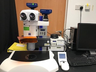
Zeiss Stereo Lumar V12 is a fully automated stereoscope with fluorescence capabilities. It allows imaging of whole small animals (e.g. Zebrafish and c. elegans), as well as cell cultures in petri dishes. Magnifications range is equivalent to ~1-12x, with variable zoom.
- Zeiss Lumar.V12 stereoscope body
Objectives
- Neo Lumar S 0.8x
Light Source
- Transmitted and reflected light source
- HBO100 fluorescence arc lamp
Filter Sets
- Zeiss Filter set 38 (GFP)
- Zeiss Filter set 20 (Rhodamine)
- Zeiss Filter set 01 (UV)
Detectors
- Zeiss AxioCam MRm
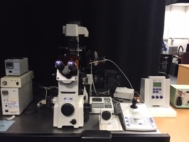
The microinjector arm module is installed on the Nikon TE2000U microscope. This system is suitable for micromanipulation and microinjection applications.
- Nikon TE2000U
Objectives
- Plan Fluor 10x/0.3
- Plan Fluor 20x/0.45
- LWD 20x/0.4
- Plan Fluor 40x/0.6
- LWD 40x/0.55
- Plan Fluor 60x/0.7
Light Source
- Mercury lamp
Excitation wavelengths
- 457nm, 488nm, 514nm
- 561nm
- 633nm
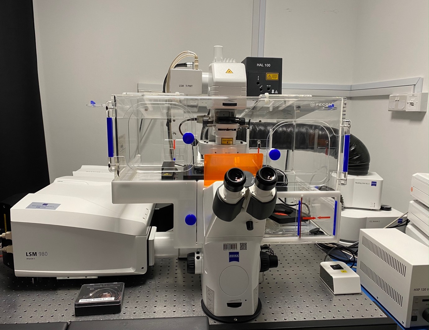
The LSM 980 Airyscan 2 is an ideal platform for fast, gentle, super-resolution imaging. It is built on a fully motorized Axio Observer 7 inverted microscope stand with Z-peizo, environmental chamber with temperature control and uses the ZEN Blue 3.4 software for acquisition and analysis. It is equipped with a 32 channel GaAsP detector, 2 PMTs and an Airyscan 2 detector. The multiplex mode can perform parallel pixel acquisition, which can accelerate acquisition speed, up to 8x faster than normal confocal imaging.
SPECIFICATIONS
Microscope body
- Axio Observer 7
- Environmental Chamber for temperature control
Objectives
- EC Plan-NeoFluar 2.5x/0.085
- Plan-Apochromat 10x/0.45
- Plan-Apochromat 25x/0.8
- Plan-Apochromat 40x/1.30 Oil DIC
- Plan-Apochromat 63x/1.40 Oil
Light Source
- Halogen lamp for transmitted light
- X-Cite 120 illuminator for reflected light
- Lasers:
- Diode Laser 405nm,445nm,488nm,514nm,639nm
- DPSS laser 561nm, 594nm
Detectors
- 32 channel GsAsp detector
- 2 standard PMTs
- Airyscan 2 detector
Software
- Zen 3.4
WORKSTATION
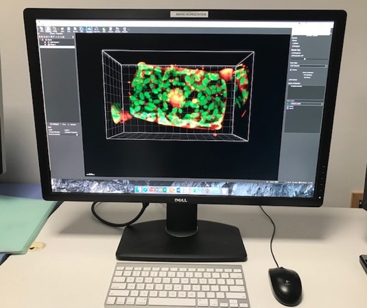 DESCRIPTION
The IMARIS single full software is installed on this workstation. This program allows the visualisation, analysis and interpretation of microscopy data. With IMARIS, you can perform automated spots and surface detection, cell segmentation, motion tracking, co-localisation and cell lineage studies etc.
