Designing a Bone Implant Around the Way Injuries Heal
NTU researchers have 3D-printed a bone scaffold designed to provide early antibacterial protection while supporting blood vessel formation and new bone growth to help repair large bone defects.
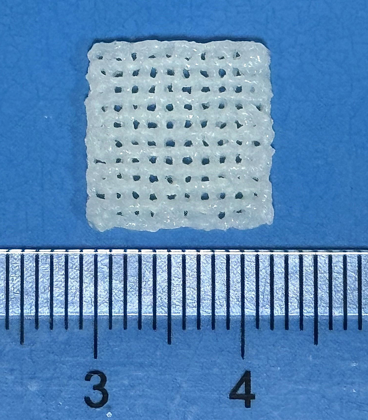 The 3D-printed scaffold is printed as a small grid a little over a centimetre across.
Its open, porous structure lets cells and blood vessels grow into it.
A small break in a bone usually mends on its own. A large one often will not.
When injury, infection, or the removal of a tumour leaves a gap too wide to bridge, the bone cannot grow back across it. The result can be lasting pain, lost movement, and repeated surgery.
For decades, the standard treatment has been to fill the gap with bone taken from elsewhere in the patient's own body.
Although effective, the bone has to be harvested in a second operation, the supply is limited, and the harvest site can develop problems of its own.
The deeper problem is that filling the defect does not guarantee successful new bone regeneration. A large defect needs to be kept free of infection, reached by new blood vessels, and then encouraged to grow fresh bone, and a transplanted block does none of that reliably.
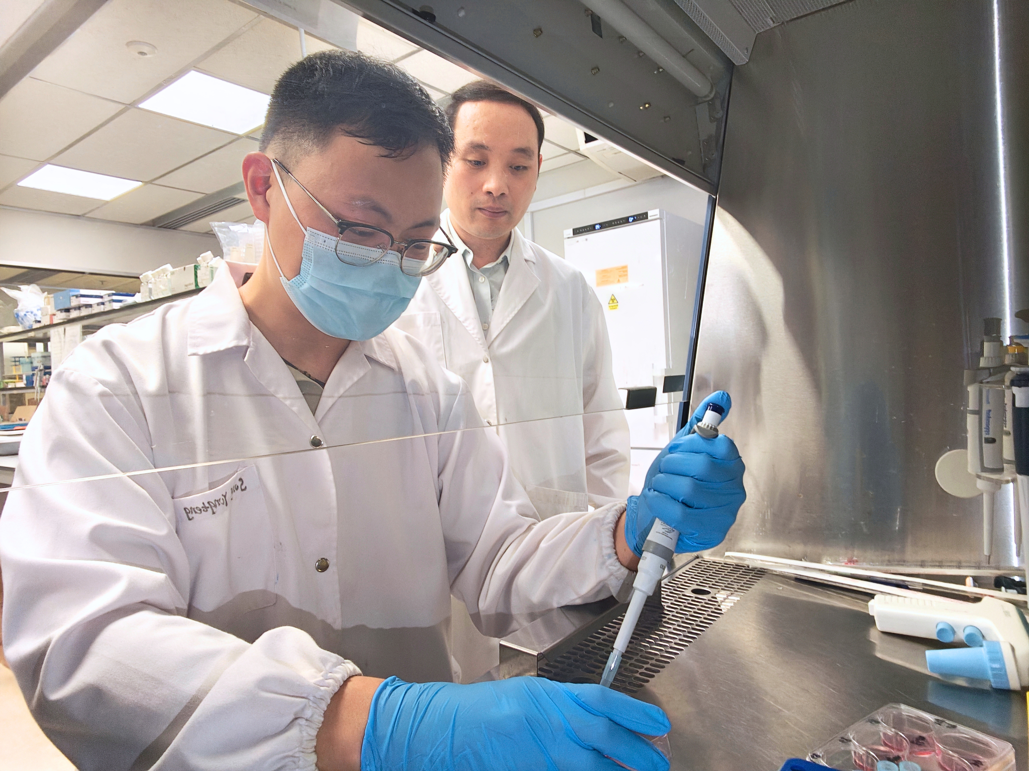 First author Yongteng Song (front) and Associate Professor Changjin Huang (behind) at work in the lab at NTU MAE.
Three problems in one wound
Healing a large bone defect means finishing three separate jobs, and they happen at different points in the process rather than all at once.
First, in the days after surgery, bacteria can settle on the wound, so it needs protection early. Then, over the following weeks, new blood vessels have to grow in to deliver oxygen and nutrients.
Only once the site is clean and fed can new bone form and mature.
Each job belongs to a different stage, which is what makes a single implant so hard to design. One that helps with a single job can easily do nothing for the other two, or arrive with the right help at the wrong time.
Loading an implant with all its medicine at once does not necessarily solve this. An infection-fighting ingredient may be released too quickly and run out too soon, and keeping some ingredients at excessively high concentrations for too long can harm the very cells healing depends on.
This is where most current approaches stop short. Antibiotic-loaded materials hold off infection but do little for blood supply, and ceramic scaffolds support bone growth but often have no defence against bacteria.
Currently, many multifunctional implants release everything at once, rather than adjusting to the wound's changing needs.
The goal is not simply to pack three functions into one implant but to have each take the lead when it is needed and then step back, without weakening the implant itself.
This is the problem Prof Huang's team set out to solve.
A release pattern that supports healing over time
The scaffold built by Prof Huang's team is printed from a soft, water-rich gel of gelatin and sodium alginate. Into that gel go two active ingredients: copper ions, and the antibiotic amoxicillin, tucked inside tiny porous hydroxyapatite ceramic beads.
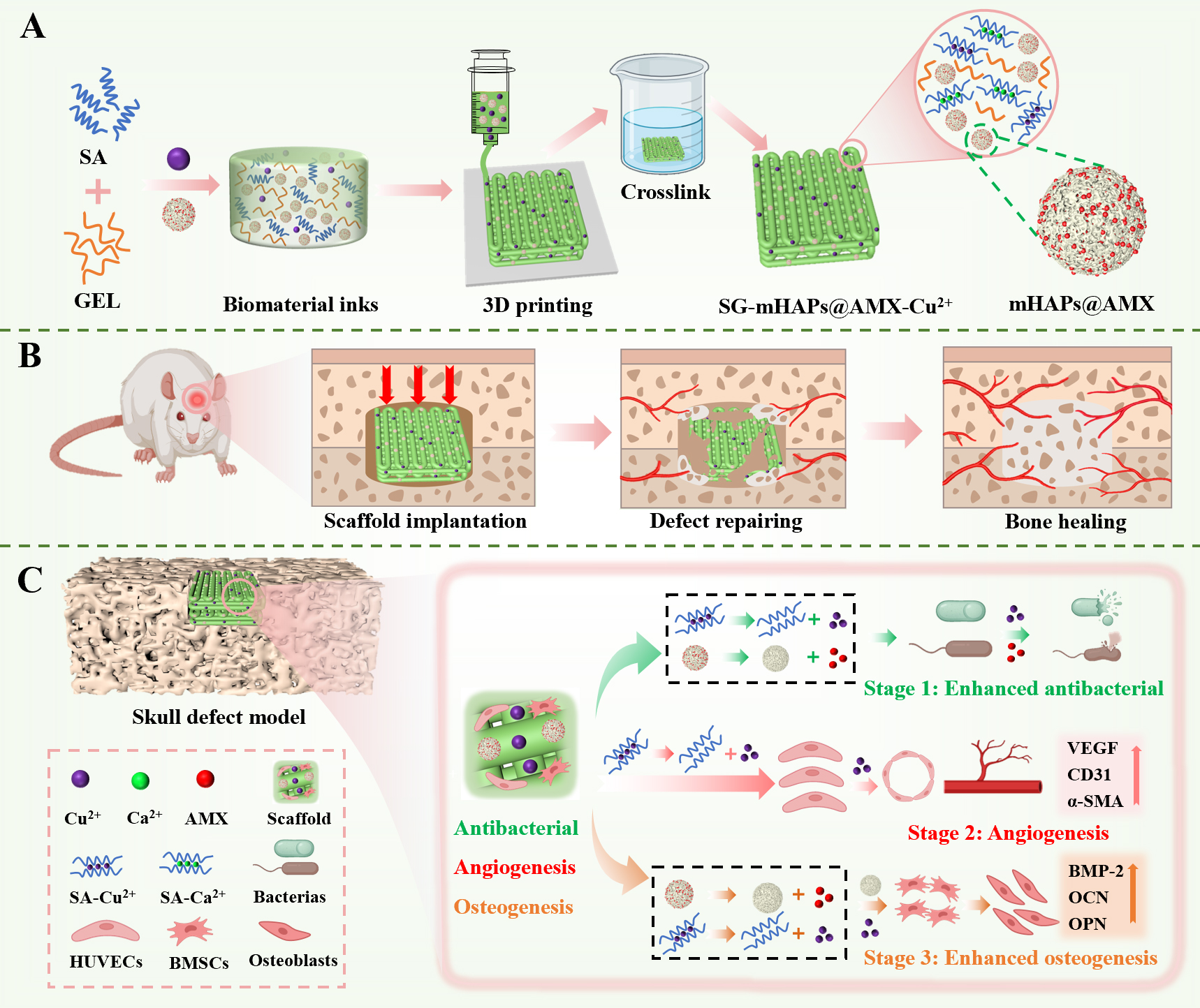 How the scaffold works. The copper ions and antibiotic-loaded microspheres are incorporated into a soft gel and 3D-printed into a scaffold (A), which is placed into the bone defect (B). Over time, the same scaffold is designed to provide early antibacterial protection while supporting new blood vessels and new bone formation (C).
The design does not switch cleanly from one treatment to the next. Both the copper ions and the antibiotic come out quickly at first, then more slowly over the following weeks.
This fast-then-slow release is the key idea.
The fast part gives strong protection at the start, when the risk of infection is highest, and the slow part keeps a steady supply going while blood vessels and bone are still forming.
The same implant covers both the early danger and the long recovery, without a second operation. What changes over time is not the ingredient but which effect matters most: first fighting infection, then feeding new blood vessels and helping bone grow.
The timing is built into how each ingredient is held.
The copper ions bind to sodium alginate within the gel network, so when the implant swells with fluid, the copper ions near the surface come out quickly first and the rest follows gradually.
The antibiotic is stored on and within the ceramic beads, each one like a microscopic sponge full of holes. The drug near the surface leaves first, while the drug deeper inside seeps out slowly through the tiny channels.
For both ingredients, the result is the same: a quick release, then a slow one.
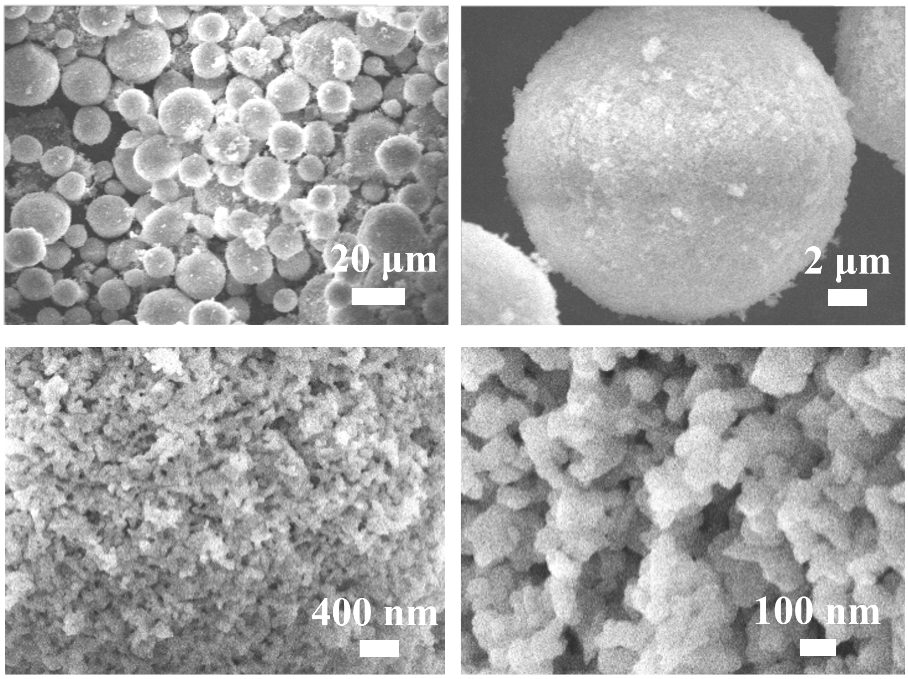 Porous ceramic beads used to carry the antibiotic, seen at different magnifications under an electron microscope.
Their sponge-like structure can hold the drug and allow it to seep out slowly.
One element doing three jobs
The most striking part of the design is how much rests on copper, an ordinary trace element the body already uses.
Copper ion fights bacteria, encourages blood vessels to form, and supports the bone-building activity of bone marrow stem cells, all while being more stable and affordable than the protein growth factors often used for the same purpose.
Each effect had been reported on its own before, Song says, but the team was still surprised that a low dose could do all three inside one printed scaffold.
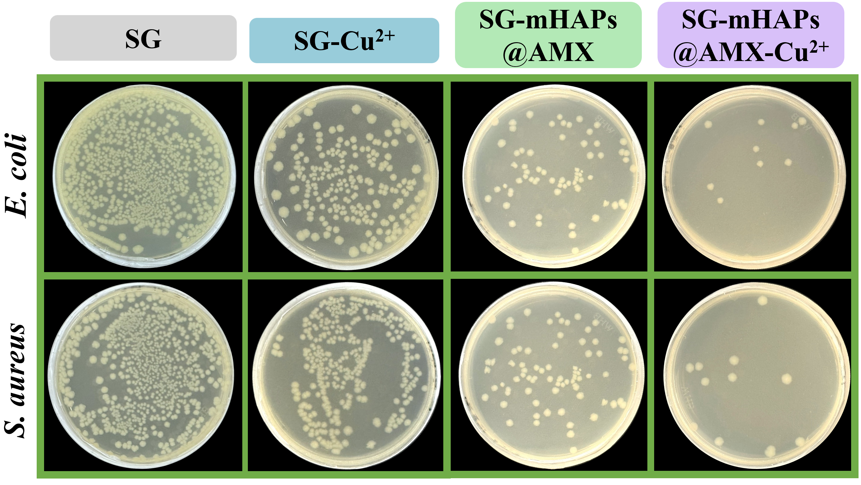 Bacterial colonies formed on agar plates after scaffold-treated bacterial suspensions were diluted and plated, with E. coli in the upper row and S. aureus in the lower row. Fewer colonies in the combined copper–antibiotic group (right) indicate stronger antibacterial activity than in the other groups.
Keeping the dose low mattered. Copper alone gave only moderate protection against bacteria, but paired with the antibiotic it produced a much stronger effect without any more copper.
The two work in different ways, which is what makes them effective together. The antibiotic stops bacteria from building their cell walls, while copper ions damage their outer membrane.
Tested against two common bacteria, the combined scaffold killed far more of them than either could alone.
What it took to build
Getting the gel to print at all was difficult. The ink had to be runny enough to flow through a fine nozzle, yet firm enough to hold its shape the moment it landed.
Too runny, and it spread into a puddle and lost its structure. Too viscous, and it clogged the nozzle or came out in broken threads.
Adding the copper ions and the beads changed how the ink behaved, so the team adjusted the formulation, temperature, extrusion pressure, and printing speed until printing became reliable.
Then came a surprise.
The porous hydroxyapatite beads were meant to carry the antibiotic and help bone form, but they also made the scaffold stronger and longer-lasting, holding its shape better and breaking down more slowly than the plain gel.
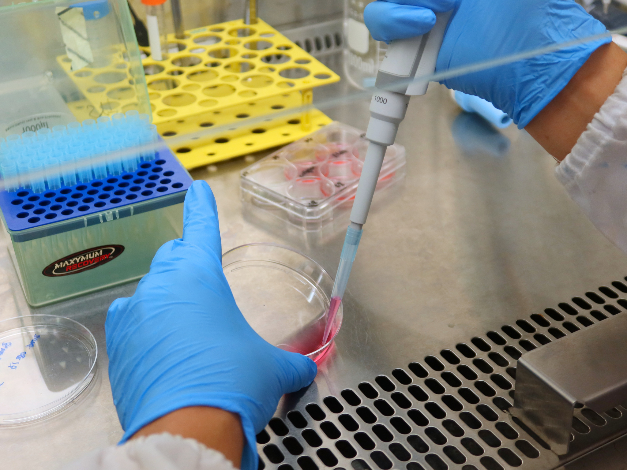 Preparing samples for cell testing in the lab. Getting the printed material to perform as intended took repeated rounds of adjustment.
The moment it came together
For Song, confidence came in two steps.
The first was in the lab, when several separate tests all pointed the same way: the full scaffold showed the strongest antibacterial activity, promoted endothelial cell migration and tube formation, and produced the highest levels of several osteogenic markers.
A single good result could have been chance, but all of them lined up with what the design was meant to do.
The second was the animal study. In rats over eight weeks, the full scaffold grew the most new bone and the most new blood vessels, with new bone extending inward from the edges of the defect toward the middle.
This was the point where the lab results and the results in a living body matched up.
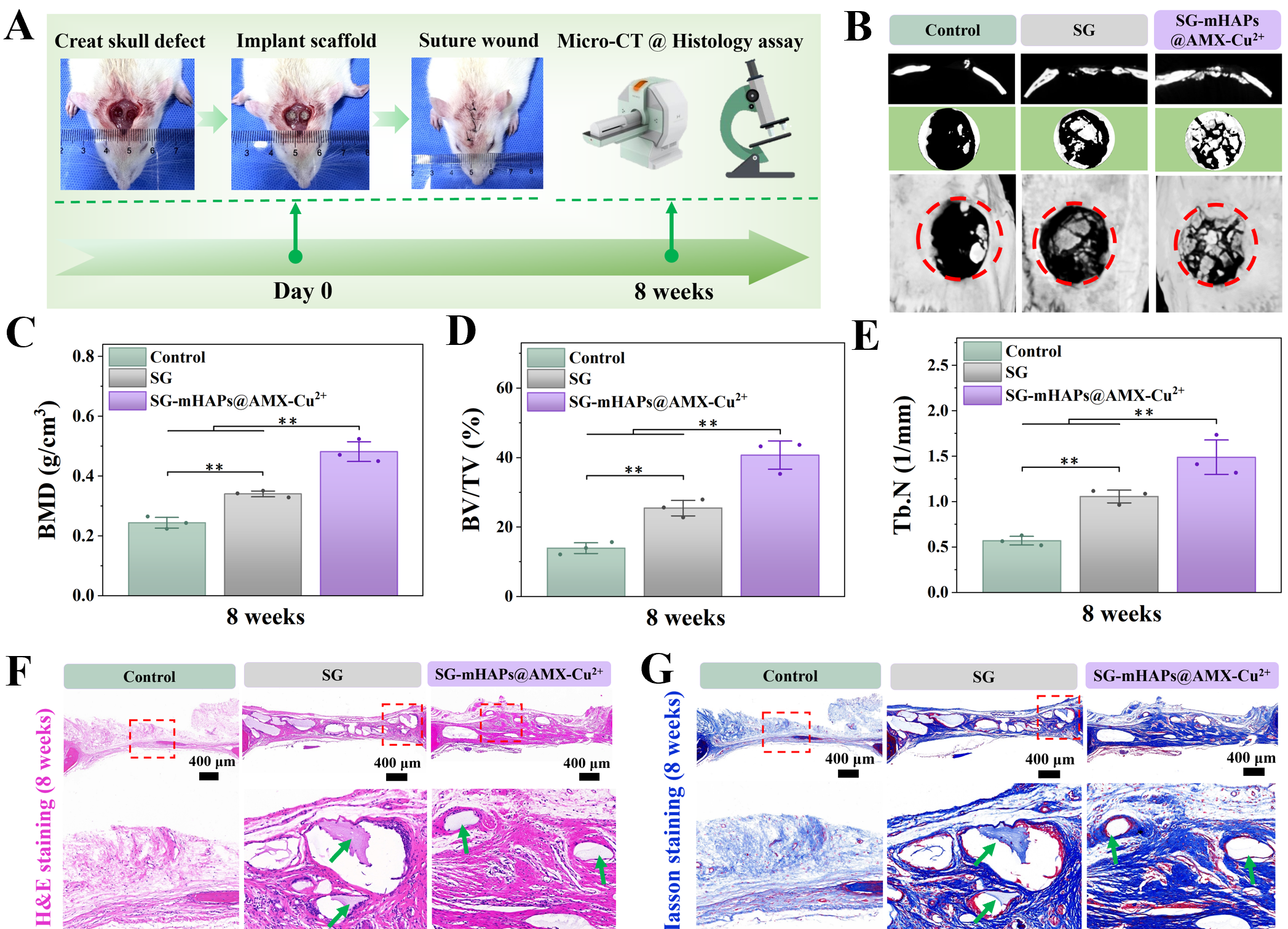 After eight weeks in a rat skull defect model, the full scaffold group (right) regenerated far more bone than an untreated defect (left), shown here in 3D micro-CT images (B) and in measurements of bone mineral density (C), bone volume fraction (D), and trabecular number (E).
An honest look at what is left to do
Song is careful about what the work does and does not yet show. It is an early proof of concept, and the eight-week test used a bone defect model without deliberately induced infection.
Before anything like it could reach a patient, it needs longer-term studies, tests in infected bone defects and larger animals, a reliable way to manufacture it, and eventually clinical trials and regulatory approval for clinical use.
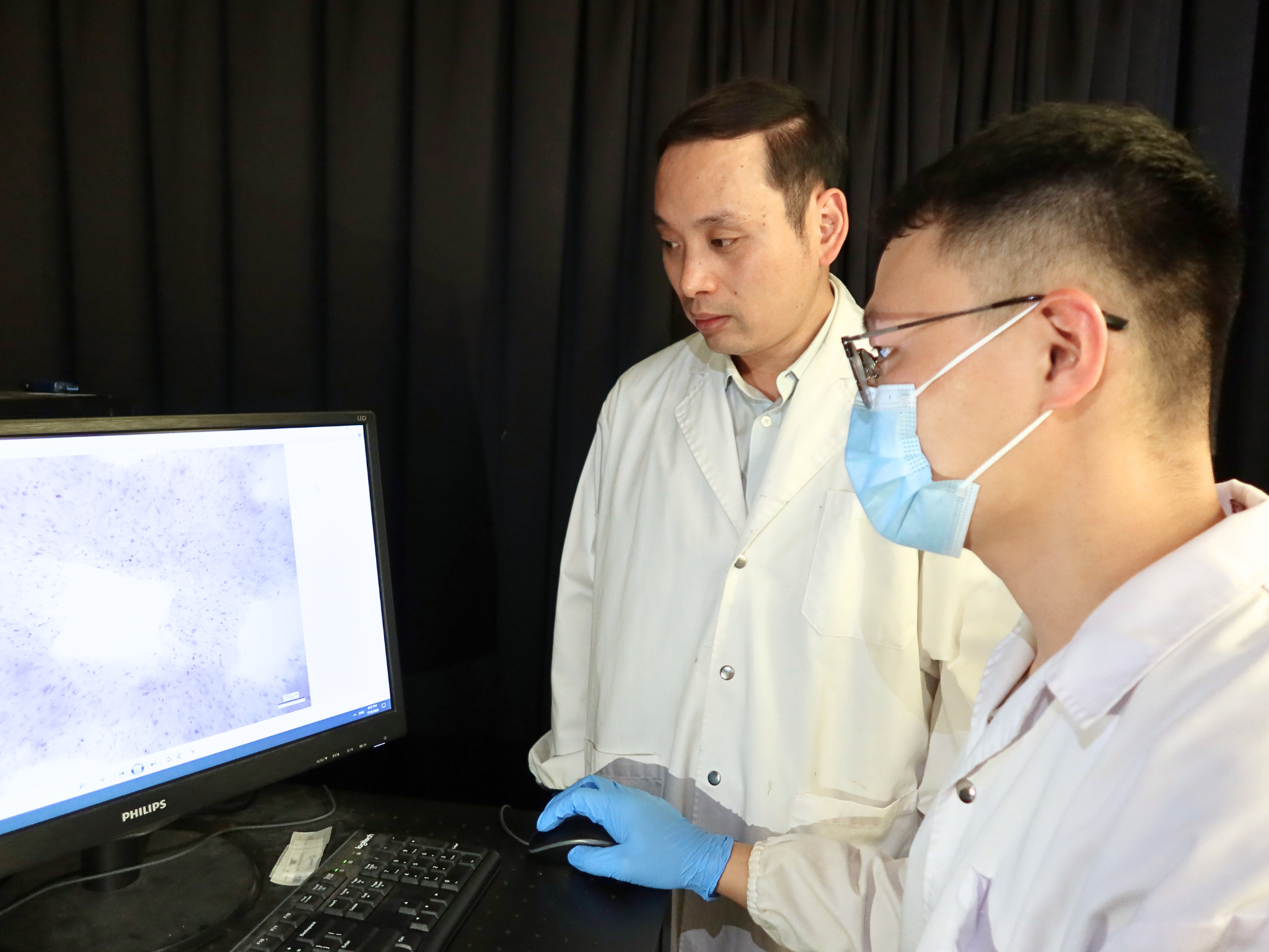 Yongteng Song and Associate Professor Changjin Huang reviewing staining results to assess the scaffold’s effects on osteogenic differentiation in the lab.
The ambition is clear, though. Song imagines the scaffold one day printed to match a patient's specific bone defect, providing early antibacterial protection while supporting blood vessel formation and bone regeneration.
For patients, that could mean more reliable healing and fewer repeat operations.
For surgeons, it could fold several treatments into a single implant, one built to do the right thing at each stage of healing.
About the research
The research was published in Materials Today Bio under the title "3D-printed multifunctional composite scaffolds incorporating copper ions/amoxicillin/hydroxyapatite for synergistic antibacterial and vascularized bone regeneration." It was supported by the National Natural Science Foundation of China, the Ministry of Education of Singapore under its Academic Research Fund Tier 1 (award RG84/25), Nanyang Technological University, and the China Scholarship Council. https://doi.org/10.1016/j.mtbio.2026.103530
About the researchers
Associate Professor Changjin Huang joined Nanyang Technological University in 2018 and holds an appointment in the School of Mechanical and Aerospace Engineering. He earned his bachelor's degree in Thermal Science and Energy Engineering from the University of Science and Technology of China, and his PhD in Engineering Science and Mechanics from Pennsylvania State University, followed by postdoctoral work at Northwestern University and Carnegie Mellon University. His research lies at the intersection of mechanics, materials, engineering, and biology, focusing on the mechanics and manufacturing of soft and living systems. |  |
Yongteng Song is the first author of the study and a researcher specialising in 3D bioprinting, biofabrication, and biomaterials. He earned his Doctor of Engineering at the Rapid Manufacturing Engineering Center in the School of Mechatronic Engineering and Automation at Shanghai University, and worked in Associate Professor Changjin Huang's group at NTU as a visiting and postdoctoral researcher. His work centres on 3D-printed tissue scaffolds and drug-loaded composite materials for bone and tissue repair. |  |
By Karen Chai, NTU MAE Communications
.tmb-listing.png?sfvrsn=892cad8a_1)