Magnetic Resonance Imaging (MRI)

Overview

Applications of MRI
MRI has a very wide range of applications in medical imaging. MRI is used to perform structural and functional imaging to understand the physiological processes of the body in both healthy and disease condition. The structural abnormalities associated with any disease condition e.g., tumor can be visualized with MRI images, which helps to perform image guided surgery. MRI is also used to perform functional imaging (fMRI) to understand the function of the brain as it responds to external stimuli and the passive activity in a resting state as well.
MRI Magnet
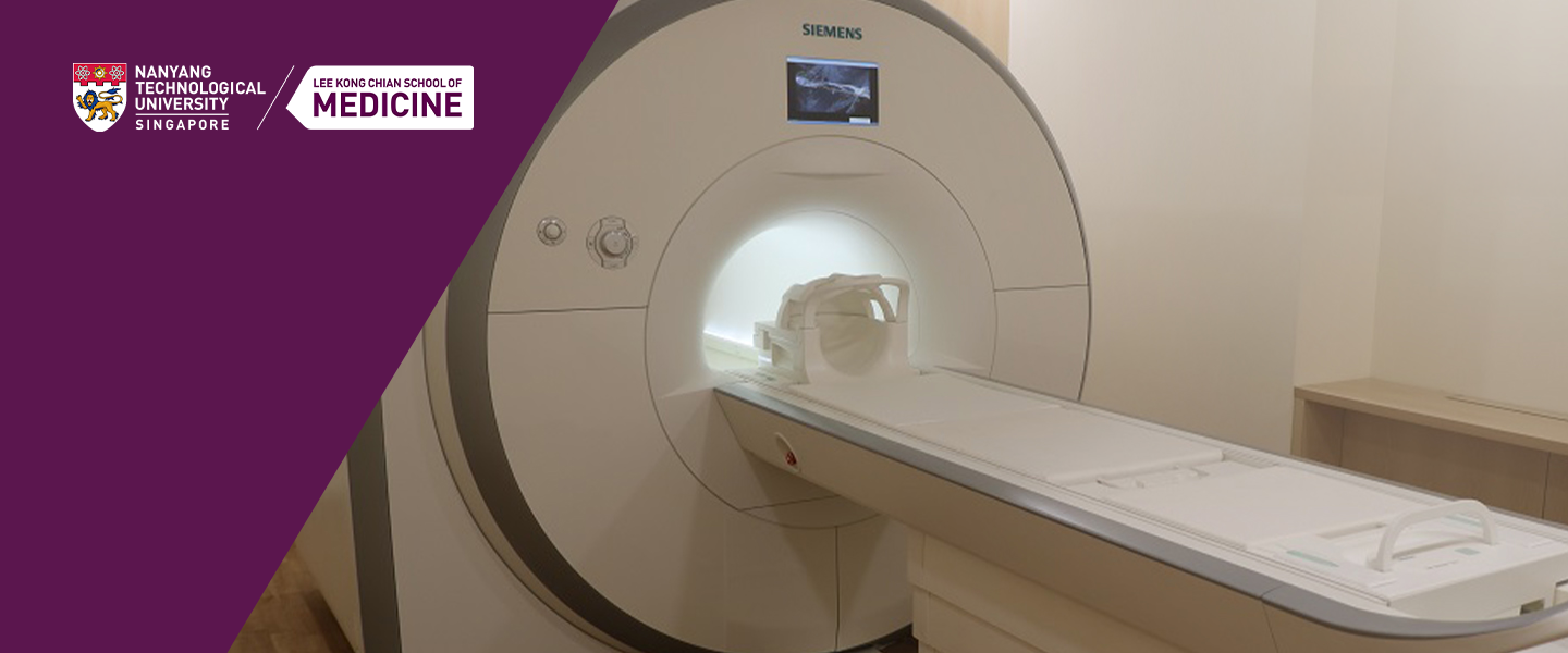
Our centre is equipped with the state-of-the-art Siemens 3Tesla PRISMA scanner with 60cm bore size. The scanner is equipped with 128 receiver channels which results in high signal-to-noise ratio images acquires with fast acquisition speed. The magnetic field stability over time is less than 0.1ppm/h. The scanner is equipped with 5th generation active shielding technology with counter coils. The External Interference Shielding (EIS) provide continuous compensation and automatic external magnetic field interferences during acquisition. The shim system comprises of three linear and five non-linear channels and a 3D patient specific automatic shimming.

Siemens 3Tesla PRISMA Scanner
RF Coils
We have a variety of RF coils suitable for imaging different parts of the body.
- Body 18
- Head 32
- Head/Neck 20
- Head/Neck 64
- Spine 32
- Flex large 4
- Flex small 4
- TxRx Knee 15
Gradient System
The scanner contains actively shielded whole-body 80mT/m gradient coil system. The gradient coils are water-cooled and with 100% duty cycle.
Methods
The scanner contains a variety of methods comprising of both spin echo (SE) and gradient echo (GE) family of sequences.
- SE
- TSE
- HASTE
- SPACE
- FLASH
- VIDE
- MEDIC
- DESS
- TurboFLASH
- FISP
- EPI
- ToF
- PC
- CV/BEAT
Patient comfort
The inner diameter of the magnet is 60cm, with in-bore lightning, ventilation and intercom.
Functional Imaging
Functional magnetic resonance imaging (fMRI), is a technique for measuring brain activity. It works by detecting the changes in blood oxygenation and flow that occur in response to neural activity – when a brain area is more active it consumes more oxygen and to meet this increased demand blood flow increases to the active area. fMRI can be used to produce activation maps showing which parts of the brain are involved in a particular mental process.
Our MRI facility contains the hardware & software for performing task based functional imaging. The fMRI kit comprises of the following:
- MR-compatible LCD monitor for visual stimuli
- MR-compatible headphones for auditory stimuli
- MR-compatible response boxes and interface
- MR-compatible eye tracking camera
- Stimulus PC
Eye Tracker
Our MRI facility have ViewPoint Eye Traker with intuitive user interface, Pupillometry, built-in state Machine for experimental control. It provides a complete eye movement evaluation, including Fixation, Saccade, drift classification, region of interest events, integrated stimulus presentation, simultaneous eye movement and pupil diameter monitoring & SDK for interfacing with other applications. Our EyeTracker system is compatible is with all the MRI head coils available in our MRI facility.
Image processing software
The data generated in scanner can be exported in DICOM format and be processed according to the user’s requirement. The Syngo.via, workstation, a Siemens post-processing software, is used for dedicated image processing purposes. In addition, the image processing is performed in 3rd party software viz.,ImageJ, Matlab, DTIStudio etc.
Data Storage & Management
The data generated per acquisition in MRI is of Gigabytes. The data generated is locally stored in the control work station, and periodically will be transferred to our Network Attached Storage (NAS). The total capacity of NAS is about 50TB. The total storage has been fragmented to about 25TB for MRI and 20TB for MEG respectively. The MRI data acquired is stored in the Siemens scanner for a brief period of time. For MRI data archiving we have Syngo.plaza storage (Siemens secondary storage system), in addition to NAS. All the storage and back-up systems are connected by local network to ensure smooth & safe data transfer and storage. The NAS has been configured (PACS) to be compatible to store the MRI data in the Siemens format. The handling of data and its delivery to the collaborators are followed according to the SOP.
Mock MRI scanner (0 Tesla)

Siemens 0Tesla Mock Scanner

Stimulation Equipment
To deliver stimulation our MRI facility is equipped with a stimulation workstation in which various built-in paradigms are present. Also the stimulation software can be used to design and develop used defined paradigms. A MRI compatible monitor behind the scanner is used to deliver the stimulation to the subject. A MRI compatible head phone is used to deliver the auditory stimulation to the subject.

Response Equipment
Two response grips are used to receive any response from the subject.
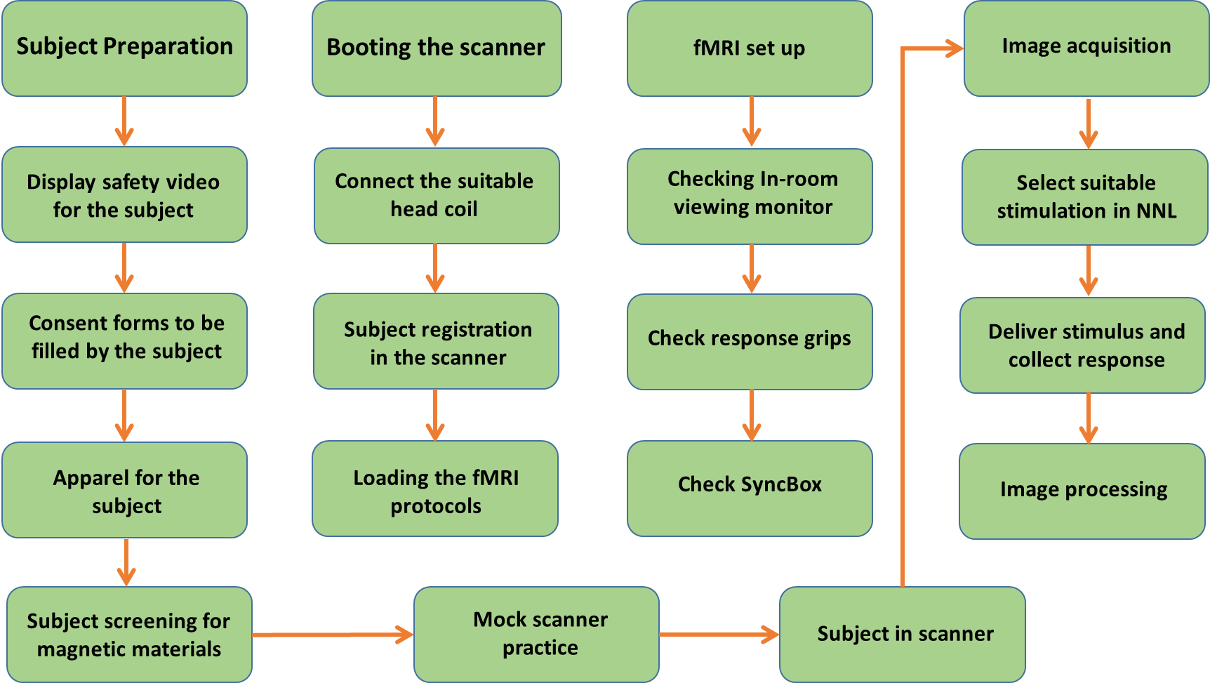
Processes


MRI: A Safe Environment
We are here to provide you the safest environment and make you aware about the potential safety hazards.
Getting Screened for Scan

Precautions for Entering MRI Scanning Room


MRI Safety Video
Videos for MRI safety and understanding the basics of MRI can be accessed from the following link provided by Siemens:
Safety Incharge
Please contact our safety in-charge regarding safety briefings and other safety issues:
1. Dr. Sundramurthy Kumar, CoNiC Safety In-charge
Email: [email protected] ; Phone: (65) - 6904 1185
2. Dr. Sini Mathew, Assistant Director, Health & Safety, LKCMedicine
Email: [email protected] ; Phone: (65) - 6592 3209

 MRI safety and understanding the basics of MRI