
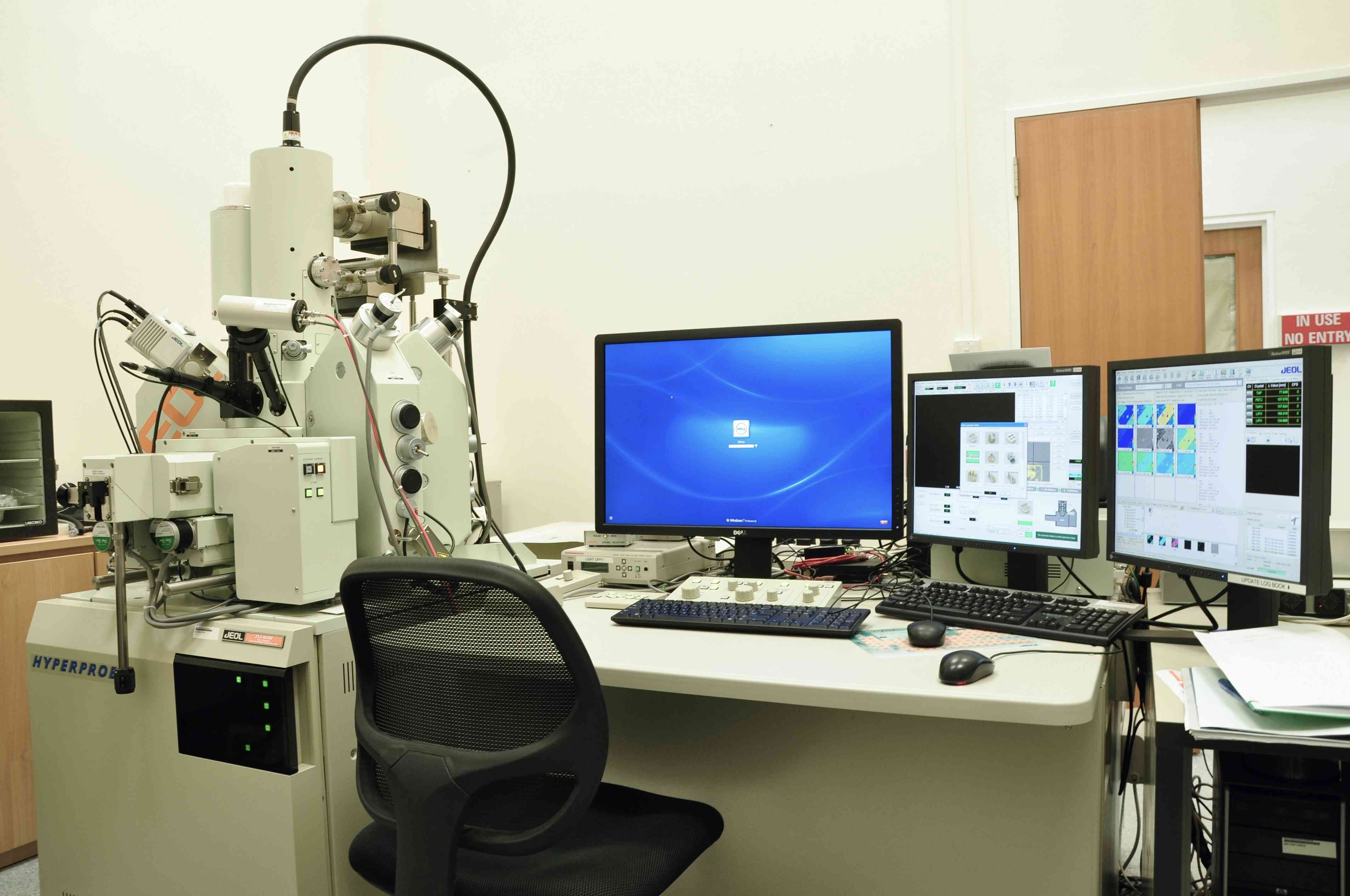
EPMA JEOL JXA-8530F
FACTS at N4.1 B4-10
Electron Probe X-ray Microanalyzer (EPMA)
The JEOL JXA-8530F field emission microprobe, installed in FACTS lab in 2010, is a precision chemical analytical tool that offers better than 1% analytical precision at spatial resolution of approximately 1-3 microns, depending upon the material. Our EPMA system is equipped with five wavelength dispersive spectrometers, an energy dispersive silicon drift detector, a secondary electron detector and a backscattered electron detector.
The electron probe X-ray microanalyzer (EPMA) is a microanalytical technique that uses the electron beam to scan the specimen and collects the emitted X-ray spectra from the sample. The use of an electron beam provides high spatial resolution (1-5 nm). The X-ray detector type is a wavelength dispersive spectrometer. Its advantages over an energy dispersive spectroscopy (EDS) detector are better spectral resolution and precision. It is typically applied to perform precision chemical analysis of metals, alloys, ceramics, minerals and glasses.
Detectable element range: WDS: Be (optional analysing crystal)/B to U, EDS: B to U
Detectable X-ray range: Detectable wavelength range with WDS: 0.087 to 9.3nm
Detectable energy range with EDS: 20keV
Number of WDS spectrometers: Up to 5 selectable; EDS spectrometers: 1
Maximum specimen size: 100 mm x 100 mm x 50 mm (H)
Accelerating voltage: 1 to 30 kV (0.1 kV steps)
Probe current range: 10-12 to 5×10⁻⁷ A
Probe current stability: ± 0.3 %/h
Secondary electron image resolution: 3 nm (W.D. 11 mm, 30 kV)
Minimum probe size: 40nm (10kV, 1×10⁻⁸ A), 100 nm (10 kV, 1×10⁻⁷ A)
Scanning magnification: x40 to x300,000 (W. D. 11 mm)
Scanning image resolution: Maximum 5,120 × 3,840
Color display:
For EPMA analysis: LCD 1,280×1,024
For SEM operation and EDS analysis: LCD 1,280×1,024
Samples can either be mounted as polished blocks ideally 25 mm (1”) in diameter or as polished sections on glass slides up to 76 x 48 mm in size (if less than 2 mm thick). Vacuum compatible epoxy resins (such as EpoFix) should be used for mounting samples. Samples must be polished to a ¼ micron diamond finish for fully quantitative analysis and, if not electrically conductive, should be carbon coated under FACTS lab supervision to ensure an even thickness consistent with coatings applied to standard reference materials used to calibrate measurements. Magnetic samples and samples that are unstable under high beam currents are not suitable for EPMA.
- X-ray microanalysis (EDX/WDX)
- Elemental composition mapping with high spatial resolution